
It is known that this is the first time this minimally invasive intervention technique has been deployed in the Mekong Delta, opening up opportunities for advanced treatment for patients with complex pancreatic diseases.
Patient PTN (30 years old, Can Tho City) was transferred to the hospital with severe abdominal pain, vomiting, diarrhea and mild abdominal distension. The patient had a history of pancreatitis for many years, poor nutrition and frequent hospitalizations.

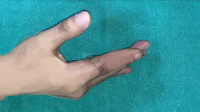
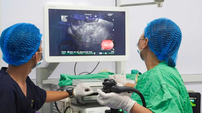
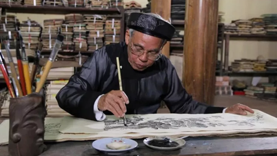
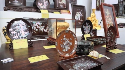
The patient's health has stabilized after the intervention. Photo: BVCC.
The results of the abdominal CT scan with contrast showed a cystic mass located next to the pancreas, behind the stomach, with signs of colon wall thickening and many dilated small bowel loops. The patient was admitted to the General Surgery Department for intensive medical treatment, to improve his condition and to evaluate the indications for intervention.
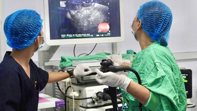
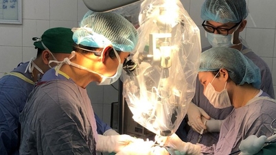
The doctor performed an endoscopy ultrasound to determine the pseudocyst of the pancreas in the tail of the pancreas, close to the stomach wall, a favorable position for drainage with a LAMS (Lumen-Appposing Metal Stent) stent, a self-expanding metal stent with a special structure, designed to create a direct communication between two adjacent organs in the body. This type of stent is widely used in endoscopy ultrasound (EUS) to drain cystic lesions or fluid-filled cavities.
After an interdisciplinary consultation, the medical team decided to intervene under the guidance of ultrasound endoscopy. During the procedure, the doctors discovered a cyst measuring approximately 9x11 cm.
Under the guidance of endoscopic ultrasound, the doctor uses a 19G aspiration needle to puncture the cyst, then inserts a guide wire into the cyst. Once the guide wire is in the correct position, the needle is withdrawn and a cutting instrument (cytotome) is passed along the guide wire to open the cyst wall.
Next, the LAMS stent is inserted through the guide wire, positioned correctly, and then expanded to create a passage between the cyst and the stomach. When the stent opens, the cyst fluid flows out, lemon yellow in color. Some of the fluid is collected and sent for testing.
As of this morning, December 9, the patient is conscious, vital signs are stable, pain has significantly reduced, abdomen is soft and no longer resistant. The patient continues to be monitored at the General Surgery Department and is expected to be discharged in the next few days.
According to Dr. Nguyen Khac Nam, Deputy Head of the Department of General Surgery, Can Tho Central General Hospital, pseudocysts of the pancreas are a common complication in patients with acute or chronic pancreatitis. If not treated promptly, the cyst can cause infection, bleeding, biliary obstruction, intestinal obstruction or rupture into the abdominal cavity. Current treatment methods include internal drainage, percutaneous drainage or surgery depending on the location, size and characteristics of the cyst wall.
Dr. Nguyen Thi Quynh Mai, Head of Endoscopy Department, Can Tho Central General Hospital, added that the LAMS stent placement technique under EUS guidance offers clear advantages: less invasive, less pain, faster recovery time and fewer complications compared to open surgery. The success of the case marks an important step forward in the diagnosis and treatment of pancreaticobiliary diseases at Can Tho Central General Hospital.
According to Dr. Mai, EUS is increasingly playing a crucial role in digestive interventions thanks to its ability to accurately identify lesions and guide safe procedures. The application of LAMS stents in pancreatic pseudocyst drainage is a modern, highly effective technique and suitable for patients with complex lesions. The successful implementation in Can Tho expands access to advanced techniques for patients in the Mekong Delta region, reducing the burden on upper-level hospitals.
Source: https://suckhoedoisong.vn/cuu-benh-nhan-viem-tuy-co-nang-lon-bang-ky-thuat-lan-dau-duoc-thuc-hien-o-dong-bang-song-cuu-long-169251209144035106.htm


![[Photo] Urgently help people soon have a place to live and stabilize their lives](/_next/image?url=https%3A%2F%2Fvphoto.vietnam.vn%2Fthumb%2F1200x675%2Fvietnam%2Fresource%2FIMAGE%2F2025%2F12%2F09%2F1765248230297_c-jpg.webp&w=3840&q=75)


![[Photo] General Secretary To Lam works with the Standing Committees of the 14th Party Congress Subcommittees](https://vphoto.vietnam.vn/thumb/402x226/vietnam/resource/IMAGE/2025/12/09/1765265023554_image.jpeg)


Comment (0)