
According to Professor, Dr. Tran Phan Chung Thuy, President of the ASEAN Otolaryngology Association, President of the Vietnam Otolaryngology Association, the nasal sinuses are located near many important anatomical structures such as the eye sockets, skull base, optic nerve, internal carotid artery...
Just one small mistake in surgery can leave serious complications such as reduced or permanent loss of vision, cerebral fluid leakage, facial paralysis, massive bleeding... due to having to operate precisely in a very small space.
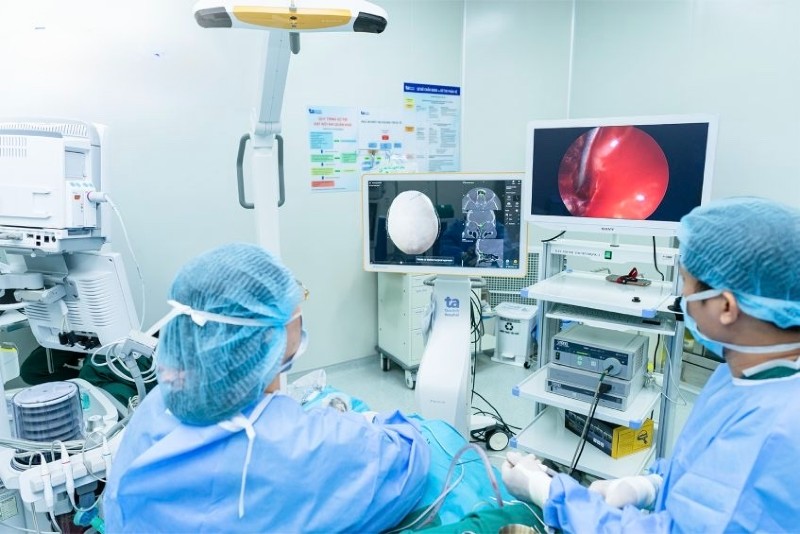 |
| Application of AI in sinus surgery at medical facilities. |
In particular, recurrent sinus surgeries are often much more complicated than the first time. At that time, the sinus structure has changed, fibrous scar tissue has formed, and natural anatomical landmarks have been lost. Determining the boundary between diseased and healthy tissue becomes more difficult, and the dissection is also more dangerous. If the inflammatory tissue is not completely removed, the disease is at risk of continuing to recur.
At the ENT Center, Tam Anh General Hospital, Ho Chi Minh City, endoscopic sinus surgery is performed with the combination of a 3D positioning system (IGS) using artificial intelligence, helping doctors proactively plan in detail before each surgery. Before surgery, the patient's CT or MRI images are uploaded to the system, AI will analyze and create a personalized anatomical map.
This system synchronizes the patient's actual anatomical data with the CT image displayed on the screen, allowing the doctor to observe simultaneously on the endoscope screen. Meanwhile, with conventional endoscopic surgery, the doctor must pause the operation to review the CT image on the display board to ensure the correct intervention position.
During surgery, the AI-integrated navigation system will provide real-time images, helping the doctor accurately determine the location that needs intervention, "guide" the doctor into the sinus, control the instrument and warn when approaching dangerous areas such as the eye socket, skull base, optic nerve... thereby minimizing complications.
“This is extremely useful for doctors in safely removing lesions, especially in recurrent sinus surgeries when the anatomical structure has changed, the risk of complications is high and absolute precision is required,” shared Professor, Dr. Chung Thuy.
This is something that traditional laparoscopic surgery cannot do, because even with the support of a camera, the surgeon's vision is still limited, especially in complex cases with scar tissue and altered anatomical structures.
Professor Chung Thuy added that the 3D positioning system using artificial intelligence also helps to perform surgery on invasive sinus tumors at the skull base more safely.
Previously, these cases required open surgery, drilling the skull first, causing craniofacial deformation and difficulty controlling tumor boundaries, with potential risk of damage to surrounding structures such as the eye socket, anterior basilar artery, optic nerve, and intracranial hematoma.
With the new system, doctors can clearly determine the location of the tumor and the surgical field limits, thereby removing the entire tumor related to the skull base and eye socket (including cancer, inverted papilloma, nasopharyngeal angiofibroma...) while still preserving the most important structures, limiting the risk of complications.
Talking about the cases she has treated, Professor, Dr. Chung Thuy said that Ms. NKP (45 years old) often had a runny nose, stuffy nose, headaches in the forehead, swelling and bulging of the left eye, and pain around the eye socket. Every time she touched her left eye socket, she felt a lump, like there was a small tumor.
Through ENT endoscopy and CT scan, Professor, Dr. Chung Thuy diagnosed Ms. P. with recurrent sinusitis, frontal sinus mucocele and deviated septum.
Frontal sinus mucocele is formed when mucus accumulates inside the sinus and cannot escape, causing blockage of the drainage duct. The tumor grows large, spreads to the eye socket, compresses the eyeball and structures around the eye, causing the patient to have bulging eyes and a heavy head in the forehead area.
Professor Chung Thuy indicated surgery to remove the inflamed tissue and mucous cyst, and to ventilate the sinuses for Ms. P. If surgery is not performed, the tumor may continue to grow, causing double vision, reduced vision, or pressure on the brain, leading to meningitis and facial paralysis.
Ms. P. had previously undergone sinus surgery to remove a frontal sinus mucocele, but the disease has now recurred. According to Professor, Dr. Chung Thuy, after the first surgery, the sinus anatomy was altered, the drainage pathways and anatomical landmarks were replaced by scar tissue, and the bone was lost, making the risk of complications if surgery continued using the traditional method very high.
This surgery was performed with the support of the Karl Storz endoscopy system (3D, 4K resolution) and a 3D AI-based navigation system. Before the surgery, Ms. P.'s CT images were imported into the system to recreate the sinus space, synchronize data and display on the screen.
After anesthesia, a sensor was attached to Ms. P's forehead. AI technology performed a 3D scan of the face, compared it with CT data, and automatically aligned anatomical landmarks, ensuring high accuracy.
During surgery, Prof. Dr. Chung Thuy uses a sensor-mounted instrument. The AI navigation system displays the instrument's path in real time on a 3D sinus map, and warns if the instrument approaches a dangerous area.
He used a specialized instrument to widen the frontal sinus drainage, creating space to access the mucocele. Under the guidance of the navigation system, the entire tumor was carefully dissected without damaging healthy tissue, while draining pus from the sinus.
The surgery was successful and finished nearly an hour earlier than expected. “I didn’t bleed as much as the previous surgery,” Ms. P. shared. After 2 days, she was discharged from the hospital and returned for a check-up a week later with good recovery.
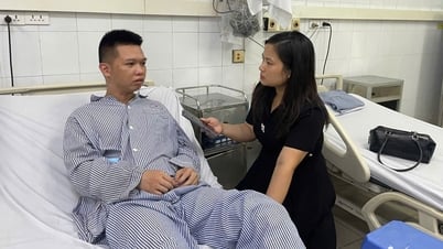
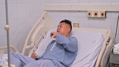
Another patient, Mr. PVT (45 years old), had chronic sinusitis and had endoscopic sinus surgery 5 years ago. However, recently, he has been experiencing continuous nasal congestion, severe headaches, and persistent purulent discharge. He was diagnosed with recurrent sinusitis, accompanied by adhesions and narrowing of the sinus drainage, a condition complicated by scar tissue formed after surgery.
According to Professor Chung Thuy, CT scans showed that Mr. T's sinus system had been altered by scar tissue, making it difficult to determine the sinus boundaries. Access to the deep lesion area using traditional endoscopy was limited.
During surgery, the AI positioning system continuously compares CT data with the patient's actual face, displaying the location of surgical instruments in real time.
This allows the surgeon to precisely identify the scar tissue that needs to be removed without damaging healthy tissue. When the surgical instrument approaches dangerous areas such as the optic nerve or the base of the skull, the AI system automatically alerts the surgeon, helping him or her make timely adjustments.
After more than an hour of surgery, all inflammation and scar tissue were removed, and the sinus drainage was cleared without causing damage to the dangerous area. “Compared to traditional methods, AI not only makes surgery safer but also shortens the surgery and recovery time for patients,” added Prof. Dr. Chung Thuy.
Source: https://baodautu.vn/tri-tue-nhan-tao-ho-tro-phau-thuat-giam-bien-chung-nguy-hiem-d324865.html


![[Photo] National Assembly Chairman Tran Thanh Man visits Vietnamese Heroic Mother Ta Thi Tran](https://vphoto.vietnam.vn/thumb/1200x675/vietnam/resource/IMAGE/2025/7/20/765c0bd057dd44ad83ab89fe0255b783)


Comment (0)