
Urothelial cancer is a type of cancer that can spread to other organs in the urinary system, including the urethra. This is an extremely rare form of cancer, accounting for only about 4%-10% of cases of recurrence after cystectomy.
Urothelial cancer is a type of cancer that can spread to other organs in the urinary system, including the urethra. This is an extremely rare form of cancer, accounting for only about 4%-10% of cases of recurrence after cystectomy.
Signs of urethral cancer
Mr. K. (72 years old, Binh Duong ) is one of the rare cases of urethral cancer, a type of cancer that accounts for less than 1% of all cancers. Previously, he had to have his entire bladder removed because of bladder urothelial cancer. However, a week ago, he discovered a sudden bleeding in his urethra and immediately went to Tam Anh General Hospital in Ho Chi Minh City for examination.
 |
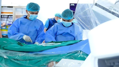
| Illustration photo |
Here, Dr. Nguyen Hoang Duc, a urologist, ordered a flexible urethral endoscopy to determine the cause. Through the endoscopy results, the doctors discovered many small tumors in Mr. K's urethra, suspected of recurrent urothelial cancer.
Urothelial cancer is a type of cancer that can spread to other organs in the urinary system, including the urethra. This is an extremely rare form of cancer, accounting for only about 4%-10% of cases of recurrence after cystectomy.
Because the malignant tumor discovered in Mr. K.'s urethra was determined to be urothelial cancer, the doctor ordered a complete urethral resection. After a day, Mr. K. recovered well, had little pain, and was able to eat and walk normally.
According to Dr. Duc, patients who have had their bladders removed and have percutaneous urinary diversion will have a reduced risk of cancer recurrence in the urethra. However, those with a history of bladder urothelial cancer are still at risk of cancer recurrence in the urethra, ureter or renal pelvis.
Doctors recommend that men and women who experience symptoms of hematuria should see a doctor immediately for timely diagnosis and treatment. Patients with a history of bladder cancer should also have regular health checks to detect early signs of recurrence.
Successful surgery for obese patient with severe osteoporosis
Mrs. Tam, 70 years old, suffered from severe pain and had to use a wheelchair for a long time. She was diagnosed with 6 damaged vertebrae due to herniated disc, severe osteoporosis and scoliosis. Although she had been treated conservatively with acupuncture and acupressure, her condition worsened, the pain spread down her legs, forcing her to stay in bed.
Ms. Tam has a BMI of 33 (severe obesity) and a bone density measurement of -3.5, which puts her in the severe osteoporosis group. According to Master, Doctor, Specialist I Vu Duc Thang, a spine specialist, surgery is the only method to help her relieve pain and restore mobility.
The surgical method indicated for Mrs. Tam was spinal screw placement to fix the vertebrae, releasing the compressed nerves and discs. The doctors also adjusted the physiological curve of the scoliosis.
However, this surgery has a high risk of complications due to the patient's severe obesity and osteoporosis. To minimize the risk, doctors used hollow screws injected with cement, which helps firmly fix the vertebrae and reduces the risk of the screws loosening or moving.
The surgery lasted 4 hours, during which 12 screws were used to fix 6 damaged vertebrae of Mrs. Tam. After the surgery, Mrs. Tam was given bone-thinning infusions to supplement calcium, vitamin D and nutrients to help strengthen bones. This method helps increase bone density and reduce the risk of fractures, at the same time she was instructed in physical therapy to restore function and muscle strength.
As a result of the surgery, Mrs. Tam no longer had any pain, gave up her wheelchair and could walk normally. The post-operative recovery period only lasted 6 days, helping her to quickly return to an independent life.
Dr. Thang shared that conservative treatment methods are always the priority, but when the disease has become severe or conservative treatment is ineffective, surgery is a necessary option to avoid serious complications. With the support of modern technology and a team of specialized doctors, spinal surgeries today bring very high efficiency."
It is known that there are modern surgical techniques for this disease, such as endoscopic surgery with biological screws, using robots to support nerve warnings and C-Arm to continuously monitor the surgical process, ensuring safety and helping patients recover quickly.
Escape the risk of complications thanks to surgery for invasive carotid tumor
A 77-year-old woman discovered a large mass in her neck after feeling a painless swelling on the left side of her neck. Initially, she thought it was a double chin due to weight gain, but after a few weeks, the mass grew larger and did not decrease. When she went to the doctor, she was diagnosed with a carotid tumor, which had surrounded the carotid artery and had begun to invade the blood vessels supplying the brain and the face and neck.
The tumor was up to 7x6 cm in size, growing rapidly from the original grape size. CT scans showed that the tumor had surrounded the carotid artery, narrowing blood flow to the brain and neck area, causing doctors to worry about the risk of serious complications if not treated promptly.
Carotid tumors are a rare type of tumor that often has no obvious symptoms in the early stages. The tumor usually develops in the common carotid artery area, where it divides into the internal carotid artery (which supplies the brain) and the external carotid artery (which supplies the face and neck). Most carotid tumors are benign, but a small percentage can be malignant.
Diagnosed with type 2 carotid tumor, the doctor determined that if left untreated, the tumor could continue to invade the entire carotid artery, even spread into the skull, causing the risk of stroke or damage to important nerves in the neck and face area.
The patient underwent tumor removal surgery under the coordination of Master, Doctor, Specialist I Le Chi Hieu and Doctor, Doctor Nguyen Anh Dung, doctors of the Department of Thoracic and Vascular Surgery.
The surgery was successful, the tumor was completely removed without causing massive bleeding or damage to important structures. Ms. Hoai recovered quickly, her chewing, swallowing, and neck and tongue movements were completely normal. After 3 days, she was discharged from the hospital, with a very low chance of recurrence thanks to the complete removal of the tumor.
Doctors advise that carotid tumors often have no obvious symptoms in the early stages. When the tumor grows large, it is easily confused with thyroid tumors or nodules. Early diagnosis through methods such as computed tomography (CT) or magnetic resonance imaging (MRI) is very important for timely treatment, avoiding serious complications.
For patients with a family history of carotid tumors, doctors recommend regular health check-ups to detect the disease early. Symptoms to watch out for include neck lumps, hoarseness, tongue numbness, sore throat, difficulty swallowing, and should seek medical attention immediately to prevent complications.
Detecting dangerous brain aneurysms from migraine symptoms
Ms. N. (65 years old, Gia Lam, Hanoi ) recently had to deal with prolonged left-sided migraines, accompanied by sleep disturbances, which made her feel anxious. Initially, she thought it was just a normal headache, but when the symptoms did not subside, she decided to go to Medlatec General Hospital for examination.
Through examination, doctors at the Neurology department suspected that she had cerebrovascular diseases, such as cerebral aneurysm or cerebral vascular malformation.
To get accurate results, the doctor ordered her to undergo a brain magnetic resonance imaging (MRI). The MRI results unexpectedly discovered a large cerebral aneurysm in the cavernous sinus of the left internal carotid artery, measuring 16mm in length, 11mm in width and 7mm in neck. Although the aneurysm had not ruptured, the doctor determined that this condition was very dangerous and required timely intervention.
Brain aneurysm is a rare condition, but extremely dangerous if not detected and treated promptly. Brain aneurysm occurs when a part of the brain artery bulges, which can compress surrounding tissues or, more dangerously, cause the artery to rupture, leading to serious complications such as stroke, coma, impaired consciousness, or death.
According to Dr. Le Quynh Son, an imaging specialist, cerebral aneurysms can be divided into three forms: saccular, fusiform, and dissecting, of which saccular cerebral aneurysms account for 85%.
Although the exact cause of the disease is unknown, some risk factors may include genetic disorders (connective tissue disease, Moyamoya syndrome, polycystic kidney disease, hyperaldosteronism), high blood pressure, smoking, estrogen deficiency in women, especially after menopause, and coarctation of the aorta.
Early diagnosis and detection of cerebral aneurysm is very important, because the disease often has no obvious symptoms in the early stages.
For early detection, magnetic resonance imaging (MRI) and computed tomography (CT) are two important methods to help determine the condition of aneurysm, predict risks and choose appropriate treatment methods.
Magnetic resonance imaging (MRI) is a safe, non-invasive and valuable method in evaluating cerebral blood vessels. Computed tomography (CT) angiography helps detect calcification or thrombosis in the arteries, thereby deciding the optimal treatment method.
Doctors recommend that people should have regular health check-ups to detect dangerous diseases such as cerebral aneurysms early. In particular, those with high risk factors, such as high blood pressure, smoking, or a family history of the disease, should pay special attention to symptoms such as severe headaches, difficulty sleeping, or sudden changes in consciousness for timely treatment.
Source: https://baodautu.vn/tin-moi-y-te-ngay-81-canh-bao-dau-hieu-ung-thu-nieu-dao-d239786.html

![[Photo] The 18th Hanoi Party Congress held a preparatory session.](https://vphoto.vietnam.vn/thumb/1200x675/vietnam/resource/IMAGE/2025/10/15/1760521600666_ndo_br_img-0801-jpg.webp)


Comment (0)