Ho Chi Minh City – Ms. Ngoc, 28 years old, frequently experiences sudden seizures. After more than 8 years of ineffective epilepsy treatment, doctors discovered she has a congenital brain defect called spina bifida.
The 3 Tesla MRI results revealed a large fissure (2-5 cm wide depending on location) in the patient's brain, extending from the cerebral cortex to the ventricle, located in the right cerebral hemisphere.
On November 10th, Dr. Chu Tan Si, Head of the Neurosurgery Department at Tam Anh General Hospital in Ho Chi Minh City, stated that the patient had suffered a brain fracture since childhood without knowing it. The fracture has now widened, causing severe epilepsy and seizures. For the past eight years, the patient has been treated for seizures and epilepsy with the highest possible dosage of medication, but without success.
Brain fissures are a congenital defect with an incidence rate of approximately 1 in 100,000 people, caused by a disorder in neuronal migration, according to Dr. Tan Si. The fissure alters cerebrospinal fluid circulation. In a normal person, cerebrospinal fluid flows from the lateral ventricles to the third and fourth ventricles, then to the periencephalic space. When a large brain fissure occurs, cerebrospinal fluid flows directly from the lateral ventricles through the fissure and into the periencephalic space, skipping several necessary stages.
According to Dr. Si, people with small brain fissures can adapt and live normally. In Ms. Ngoc's case, the fissure widened over time, allowing cerebrospinal fluid to leak in, increasing intracranial pressure and compressing the surface of the cerebral cortex, causing epileptic seizures. The optimal solution is decompression surgery, followed by continued monitoring and treatment of epilepsy.
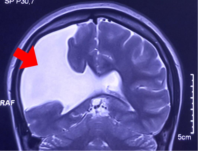
A cranial MRI scan revealed a large brain fissure with cerebrospinal fluid leakage (white area). (Image: Provided by the hospital )


The surgeon performs intracranial decompression, inserting a specialized plastic tube into the brain fissure and down into the peritoneal cavity. The tube is equipped with an automatic valve that maintains a moderate cerebrospinal fluid (CSF) level, stabilizing intracranial pressure. When CSF levels at the brain fissure increase, raising intracranial pressure, the tube automatically opens, allowing CSF to flow into the peritoneal cavity. When CSF levels decrease, the valve automatically closes, preventing the CSF from dropping too low.
Three days after surgery, the patient's health is stable, recovering well, and is expected to be discharged in 5 days.
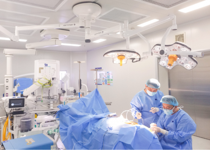
Surgeons perform brain decompression surgery on a patient. Photo: Provided by the hospital .
Dr. Tan Si stated that patients need to take anti-epileptic medication at appropriate dosages. Doctors monitor changes in cerebrospinal fluid and intracranial pressure, as well as epileptic seizures, for 2-6 months and adjust the anti-epileptic medication prescription accordingly. The goal is to transition from multi-drug therapy to monotherapy, from the highest possible dose of anti-epileptic medication to the lowest possible dose, helping patients gradually improve their health and quality of life.


Truong Giang
* The patient's name has been changed.
| Readers can post questions about neurological diseases here for doctors to answer. |
Source link