

Post-cesarean section wound fluid accumulation is a fairly common condition today, causing mothers to face many health problems such as prolonged menstruation, vaginal bleeding, infection… and even the risk of secondary infertility. To update professional knowledge on this issue and provide valuable discussions from an expert's perspective, the MEDLATEC Healthcare System's professional development program, week 40, with the theme "Approach and Management of Post-cesarean Section Wound Fluid Accumulation," was successfully organized.
The MEDLATEC Healthcare System's professional development program for week 40 was chaired by Assoc. Prof. Dr. Nguyen Quoc Dung – Vice Chairman of the Science, Technology and Training Council; Director of Professional Services at the MEDLATEC Diagnostic Imaging Center. In addition, the program was attended by Assoc. Prof. Dr. Dang Thi Minh Nguyet – Obstetrics and Gynecology Specialist, MEDLATEC Healthcare System, Deputy Head of the Obstetrics and Gynecology Department – Hanoi Medical University; Deputy Head of the Gynecological and Endocrinology Department – National Obstetrics and Gynecology Hospital, along with a team of experienced speakers and a large number of doctors and medical staff from across the system.
Signs and examination process for cases of effusion at a previous cesarean section wound.
To begin the program, Dr. Nguyen Thi Thuy Linh, Specialist in Obstetrics and Gynecology at MEDLATEC General Hospital, presented an overview of a real-life clinical case that was successfully treated at the unit.
For the past year, after her menstrual cycle ends, Ms. TTQ (31 years old, Ha Tinh ) has often had a brown discharge lasting about a week. After giving birth to her first child in 2018, she and her husband have been hoping for another child for the past year.
For those reasons, Ms. Q. went to MEDLATEC General Hospital for examination. After reviewing the patient's and family's medical history, the doctor found no abnormalities. Ms. Q. was prescribed necessary paraclinical tests to aid in diagnosis.
The ultrasound images showed fluid accumulation at the site of the previous cesarean section scar, measuring 7.4 x 6.8 mm. Transvaginal ultrasound revealed a fluid-filled cavity measuring 17 x 6 mm in the isthmus region, with 2.5 mm of muscle remaining. Based on this, the doctor concluded that the patient had fluid accumulation at the site of the previous cesarean section scar and advised treatment using hysteroscopy for scar revision.
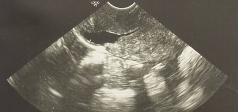
Ultrasound images showed fluid accumulation at the site of the previous cesarean section scar, measuring 7.4 x 6.8 mm.
Based on the presentation of clinical case information, Dr. Nguyen Thuy Linh emphasized the key points to note during the diagnosis and treatment of this condition to all attending medical professionals.
From A to Z: Information about open cesarean section scars
Next, MSc. Dr. Ha Ngoc Manh – Deputy Director – Head of the Department of Andrology, Vietnam-Belgium Specialized Hospital for Andrology and Infertility, presented the second report on the topic "Cesarean section scar dehiscence – diagnosis and treatment".
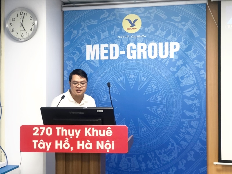
MSc. Dr. Ha Ngoc Manh presented a report on the topic "Cesarean section scar dehiscence – diagnosis and treatment".
Within the framework of this report, valuable general knowledge on this issue has been clarified by experts, including:
1/ Definition:
A cesarean section scar dehiscence is defined as:
- Secondary cavities (spaces) appear after cesarean section, located in the anterior wall of the uterine isthmus;
- It may contain fluid, which could be menstrual fluid or secretions causing chronic inflammation;
- Formed due to abnormal scar healing;
- The structure is a cavity, the inner surface of which is lined with mucous membrane.
2/ Classification:
Based on the volume of the scar tissue, cesarean section scar dehiscence is classified as follows:
- Grade 1: Less than 15 cubic millimeters;
- Grade 2: 16 – 25 cubic mm;
- Grade 3: Over 25 cubic millimeters.
3. Complications:
- Secondary infertility;
- During pregnancy;
- Pregnancy at the cesarean section scar;
- Placenta previa at the incision site;
- Spinach;
- Young water spinach;
- Uterine rupture;
- Postmenstrual bleeding;
- Menstrual cramps, pelvic pain, pain during sexual intercourse;
- Uterine-bladder fistula.
4/ Diagnosis:
Clinically, open wounds from previous surgical scars can be identified through several signs, including the following:
- Abnormal uterine bleeding;
- Brown or black bleeding a few days after menstruation;
- Pelvic pain/menstrual cramps;
- Secondary infertility.
For paraclinical examinations, imaging techniques play a crucial role in diagnosing open wounds from previous surgeries, including:
- Supersonic;
- Ultrasonic water pump;
- Hysterosalpingography (HSG)
- Magnetic resonance imaging (MRI).
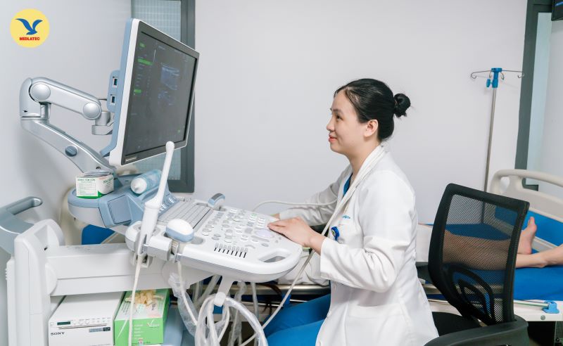
Diagnostic imaging techniques have important applications in diagnosing open wounds from previous surgical incisions.
In particular, experts emphasize that the indicators that need to be measured on ultrasound/MRI include:
- Depth of scar tissue opening;
- Length of the scar tissue opening;
- The remaining uterine muscle layer;
- Adjacent uterine muscle layer;
- Endometrial thickness and endometrial fluid layer;
- Width of the scar tissue opening.
5/ Treatment:
Treatment is individualized for each patient. Accordingly, the clinician will take a medical history, perform a physical examination, and order necessary diagnostic tests. Based on the diagnostic results, they will prescribe a suitable treatment plan that is highly effective and meets the patient's wishes.
Common methods include:
– Medical treatment: Oral contraceptives (applied to patients who do not wish to have children).
– Surgical treatment:
- Laparoscopic abdominal surgery;
- Vaginal surgery;
- Hysteroscopy.
Among the methods presented, MSc. Dr. Ha Ngoc Manh emphasized that hysteroscopy is considered the first-line treatment option due to its simplicity and effectiveness.
With a spirit of seriousness and urgency, the professional development program at the MEDLATEC Healthcare System, themed "Approach and management of fluid accumulation in old cesarean section wounds," concluded successfully.
The program was lively with exchanges and discussions, which helped the medical staff acquire useful knowledge and apply it effectively in their practice of diagnosis and treatment.
On its journey to fulfill its mission of caring for community health, MEDLATEC continuously strives to improve its professional capabilities and pioneer the application of advanced techniques from the world's medical field to further enhance the quality of diagnosis and treatment for the people.
With the advantage of gathering a team of leading experts and doctors along with a system of advanced and modern equipment, the MEDLATEC Healthcare System accurately diagnoses the condition of old surgical scar open wounds, thereby guiding appropriate treatment plans and maximizing effectiveness for patients. For in-depth consultation or to schedule an appointment, please contact the hotline at 1900 56 56 56, available 24/7. |
Source: https://medlatec.vn/tin-uc/tiep-can-va-xu-tri-benh-ly-u-dich-vet-mo-de-cu



![[Image] Close-up view of the interchange connecting the two expressways and Long Thanh Airport.](https://vphoto.vietnam.vn/thumb/1200x675/vietnam/resource/IMAGE/2026/05/25/1779703378210_ndo_br_z7863716673926-224453a31600126cce10622af6290afd-4549-jpg.webp)









































































Comment (0)