

Dr. Nguyen Dinh Lien, from the Department of Urology and Andrology at E Hospital, said that the 65-year-old patient was admitted with severe bilateral lower back pain and prolonged fatigue due to kidney stones and ureteral stones causing urinary tract obstruction.
Spinal rigidity for 18 years
Imaging results confirmed the patient had hydronephrosis caused by obstructive kidney stones. However, what made the case particularly complicated was the severe scoliosis that had persisted for many years.
According to the patient's medical history, he was involved in a traffic accident nearly 20 years ago. After the accident, his spine gradually became severely deformed, his body increasingly bent over, and he lost the ability to move normally. For 18 years, he lived with almost constant pain, experiencing difficulties in daily life and a noticeable decline in his physical condition.
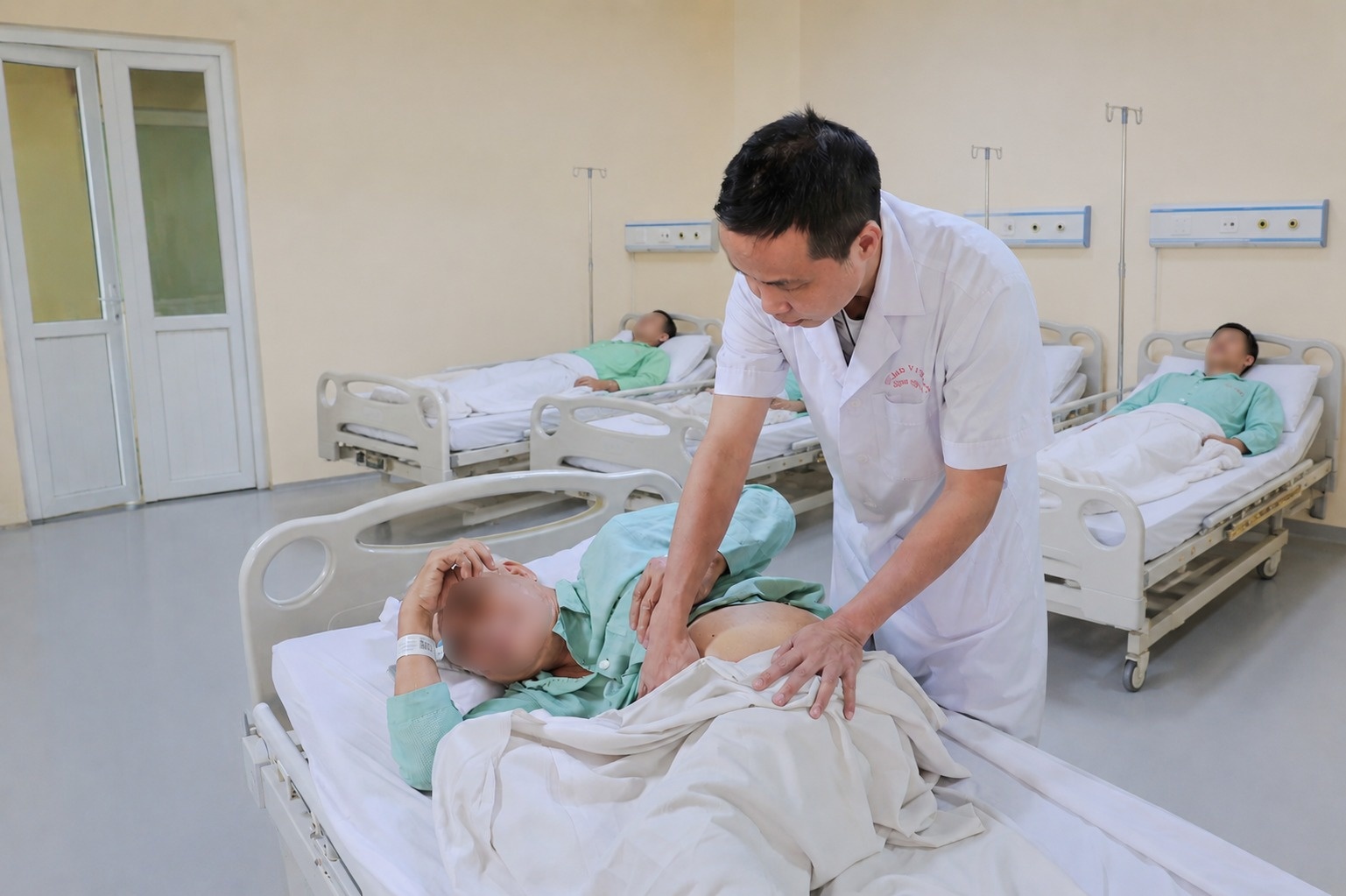 |
A doctor examines a 65-year-old patient. Photo: Provided by the hospital. |
About a year ago, the patient was diagnosed with kidney stones. His family took him to several medical facilities, but they all faced difficulties in treatment due to his advanced age, weak physical condition, complex spinal deformity, and multiple underlying health conditions.
"The patient has a very severe spinal deformity, making it almost impossible to maintain a normal lying position. This is a factor that makes both anesthesia and surgery potentially risky," shared Dr. Nguyen Dinh Lien.
Special anesthesia procedure when the patient cannot lie flat.
According to the medical team, the first difficulty arose right from the anesthesia and resuscitation stage.
Normally, patients are placed supine for endotracheal intubation for anesthesia. However, in this case, ankylosing spondylitis caused the cervical and lumbar spine to lose almost all mobility. The patient could not lie flat and could only maintain a fixed, flexed position like a "shrimp."
This makes airway management particularly challenging. Anesthesiologists must use a flexible endoscope to intubate – a technique typically reserved for complex airway cases, requiring considerable experience and close coordination among team members.
After successfully controlling anesthesia, another challenge arose right on the operating table.
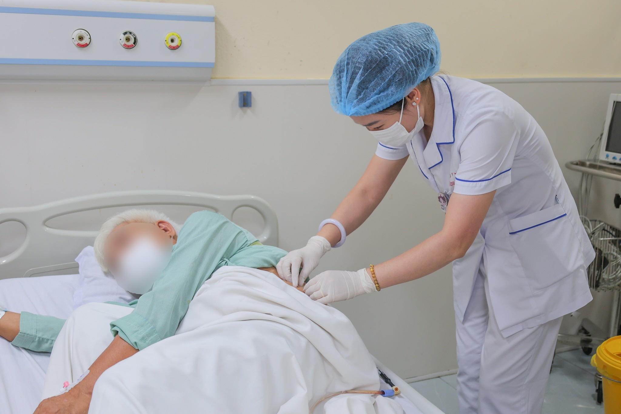 |
This is a very rare case because the patient's anatomy is almost completely altered. Photo: Provided by the hospital. |
Initially, doctors planned to use retrograde endoscopy to access and treat the ureteral stones. This is a minimally invasive technique, often preferred in the treatment of urinary tract stones.
However, during the procedure, the team found that the patient's ureter had become severely deformed due to prolonged curvature. The abnormally angled ureter made it extremely difficult to access the stone with instruments.
Right in the operating room, under the direction of Dr. Nguyen Dinh Lien, the surgeons quickly consulted and decided to change the strategy, switching to percutaneous endoscopic lithotripsy.
While this might seem like a more favorable approach, difficulties continued to arise. Due to anatomical deformities that had persisted for many years, the patient's kidney was located very high up, close to the rib cage. The narrow, constricted ribs severely limited access to the renal pelvis.
"This is a very rare case because the patient's anatomy is almost completely altered. Every step requires extremely precise calculations to find a safe access route to the renal pelvis," said Dr. Nguyen Dinh Lien.
According to doctors, even a slight deviation during the puncture process can put the patient at risk of pleural damage, bleeding, or failed access.
After many efforts, the team finally created a safe access route to the renal pelvis. In just about 10 minutes of lithotripsy, the rough-surfaced, firmly attached ureteral stone in the upper third of the ureter, which had been causing a long-term obstruction, was successfully removed, freeing the urinary tract that had been blocked for an extended period.
According to Doctor Lien, if left untreated, prolonged obstruction can severely impair kidney function, increase the risk of urinary tract infections, and lead to many other dangerous complications.
Thanks to the minimally invasive treatment method, patients experience significantly less post-operative pain, recover faster, and have a reduced risk of impaired kidney function.
The expert also advised people not to be complacent about symptoms such as lower back pain, painful urination, difficulty urinating, or persistent pain in the hip and lower back area, as these could be signs of urinary tract stones. Early examination helps in timely detection and treatment, avoiding the risk of dangerous complications affecting kidney function.
Source: https://znews.vn/than-chui-len-nguc-bac-si-mo-duong-cuu-ca-kho-hiem-gap-post1654971.html
![[Photo] Standing Committee member of the Party Central Committee Tran Cam Tu working with the Central Inspection Committee](https://vphoto.vietnam.vn/thumb/1200x675/vietnam/resource/IMAGE/2026/05/28/1779969579668_ndo_br_bnd-2495-jpg.webp)













































































Comment (0)