

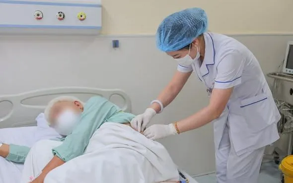
Photo: BVCC
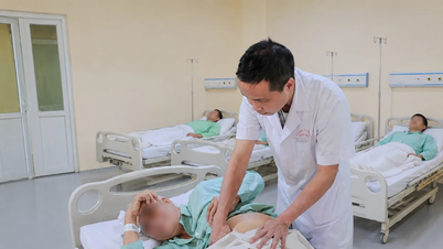
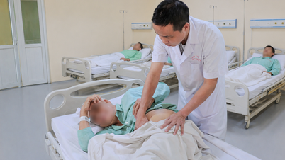
The patient was admitted with severe bilateral lower back pain and persistent fatigue due to kidney and ureteral stones causing urinary tract obstruction. Examination and imaging revealed hydronephrosis caused by the stones. However, what made the case particularly complex was the patient's long-standing ankylosing spondylitis, which had left the body almost completely rigid and unable to lie flat.
Dr. Nguyen Dinh Lien, Head of the Urology and Andrology Department at E Hospital, said that the patient had been in a traffic accident nearly 20 years ago. After the accident, his spine was severely deformed, his body increasingly bent over, he lost the ability to move normally, and he had to live with prolonged pain. Nearly a year ago, the patient was diagnosed with kidney stones, but many medical facilities were hesitant to intervene due to his advanced age, weak physical condition, and the overly complex anatomical deformity.
Difficulties arose right from the anesthesia and resuscitation stage. Because the patient could not lie supine, doctors had to use flexible endotracheal intubation – a method reserved for cases with difficult airways, requiring considerable experience and close coordination between specialties.
Initially, the team planned to perform retrograde endoscopy to treat the ureteral stone. However, due to the abnormal angle of the ureter caused by prolonged spinal curvature, the instruments had difficulty accessing the stone. Right in the operating room, the doctors decided to switch to percutaneous lithotripsy.
However, the challenge continued when the patient's kidney was located very high up, close to the rib cage, and the ribs were tightly packed, severely limiting access to the renal pelvis. Even a small mistake during the puncture process could cause damage to the pleura or bleeding.
"This is a rare case because the anatomy is almost completely altered. Every step requires extremely precise calculations to find a safe access route to the renal pelvis," shared Dr. Nguyen Dinh Lien.
After much effort, the team successfully created a safe access route to the renal pelvis and removed the long-standing obstructing ureteral stone in the upper third of the ureter. Thanks to the minimally invasive technique, the patient experienced reduced postoperative pain, faster recovery, and minimized the risk of impaired kidney function.
Doctors advise people not to ignore symptoms such as lower back pain, painful urination, difficulty urinating, or persistent pain in the flank area, as these could be signs of urinary stones. Early detection and treatment help reduce the risk of kidney dysfunction and dangerous complications.
Source: https://vtv.vn/than-chui-len-nguc-cot-song-gap-cung-van-duoc-tan-soi-thanh-cong-100260528230020602.htm



![[Image] Nearly 125,000 Hanoi students completed the Literature exam for the 10th grade entrance exam.](https://vphoto.vietnam.vn/thumb/1200x675/vietnam/resource/IMAGE/2026/05/30/1780117233041_ndo_br_thiet-ke-chua-co-ten-2-png.webp)












![[Video] Over 5,700 establishments found in violation, fined over 22.4 billion VND during peak month of food safety inspection.](https://vphoto.vietnam.vn/thumb/402x226/vietnam/resource/IMAGE/2026/05/30/1780130426976_dung00-12-11-08still005-png.webp)






































































Comment (0)