Facing dangerous coronary artery restenosis because of ignoring warning signs
Mr. Trong, 78 years old, was diagnosed with high blood pressure, diabetes and a history of myocardial infarction and had a coronary artery stent placed in 2010. Since then, he has been taking medication regularly as prescribed.
"For the past 3-4 years, I have occasionally had chest pain that lasted for a few minutes and then went away on its own. I thought it was due to old age. The doctor advised me to have my coronary arteries checked again because I had a stent placed a long time ago, but because I was subjective, I have not had it checked again," Mr. Trong shared.
A few days before being admitted to the hospital, Mr. Trong experienced chest pain, which tended to last longer and become more frequent with exertion. Sometimes he felt a heavy pressure on his chest.
"Through clinical examination and cardiac enzyme test index (Troponin T) with a slight increase, we suspect that Mr. Trong has progressive coronary artery disease after 15 years of stent placement, so we ordered percutaneous coronary angiography for examination," said Master, Doctor Cao Manh Hung - Department of Cardiology - Interventional Cardiology, Hong Ngoc General Hospital.
Coronary angiography images showed that Mr. Trong had severe restenosis in the stent. This is a phenomenon in which the coronary artery narrows again at the site where the stent was placed, causing myocardial ischemia, a fairly common problem in long-term follow-up after intervention. Restenosis can appear after 6 - 12 months after intervention, sometimes longer as in Mr. Trong's case, after nearly 20 years.
In addition to severe calcified atherosclerotic lesions causing tight stenosis (> 95%) in the old stent at segment II, angiography results also recorded diffuse calcified lesions causing about 80% stenosis in the proximal segment of the anterior interventricular artery - the largest coronary artery branch supplying the heart.
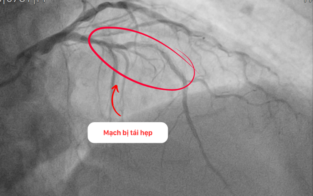
The artery is re-narrowed.
Complex stent restenosis intervention with special cutting balloon
BSCKII. BSNT Le Duc Hiep - Cardiology - Interventional Cardiology Department of Hong Ngoc General Hospital, who directly participated in the intervention for Mr. Trong, said that the artery supplying Mr. Trong's heart was almost completely narrowed - the cause of the patient's angina attacks. This caused a serious shortage of blood supply to the heart, which could lead to myocardial infarctions, threatening life at any time if early intervention to re-open the coronary arteries was not performed.
The team decided to perform coronary angioplasty and stent placement in segments I and II of the anterior interventricular artery - the main culprit causing angina attacks - to ensure recovery of myocardial contractility and improve Mr. Trong's symptoms.
The biggest difficulty in this intervention was severe restenosis in the stent with high levels of calcification and fibrosis, making it difficult to widen the lumen of the vessel at the site of strict stenosis.
"If dilating with a conventional balloon, it is very easy for the balloon to slip, or not widen the vessel. In that case, the doctor must pump at very high pressure, increasing the risk of damage to the vessel wall, causing many complications and a very high possibility of re-narrowing," said Dr. Hiep.
After a thorough consultation, the intervention team decided to use a special device called a cutting balloon. This type of balloon has small blades attached to its body to help cut and destroy calcified atherosclerotic lesions with moderate pressure to effectively expand the lumen of the blood vessels.
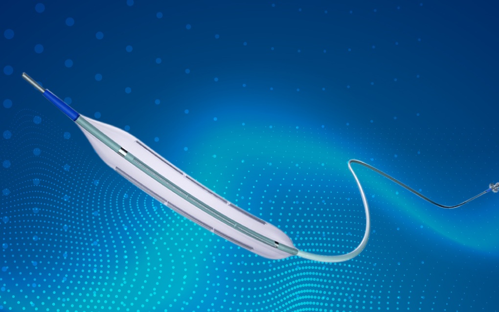
Illustration of Cutting Balloon (Photo: Lepu Medical).
After gradually treating the injury, the team successfully inserted a stent to prevent restenosis.
With the support of the IVUS system, the team accurately measured the size of the vascular lumen, the length of the lesion, and the appropriate location for stent placement. From there, they selected the balloon and stent with the appropriate size to ensure patient safety and optimize intervention effectiveness.
After 60 minutes of careful consideration, the team successfully placed two stents into segments I and II of the anterior interventricular artery. Intravascular ultrasound after the procedure showed that the stent expanded well and was close to the vessel wall.
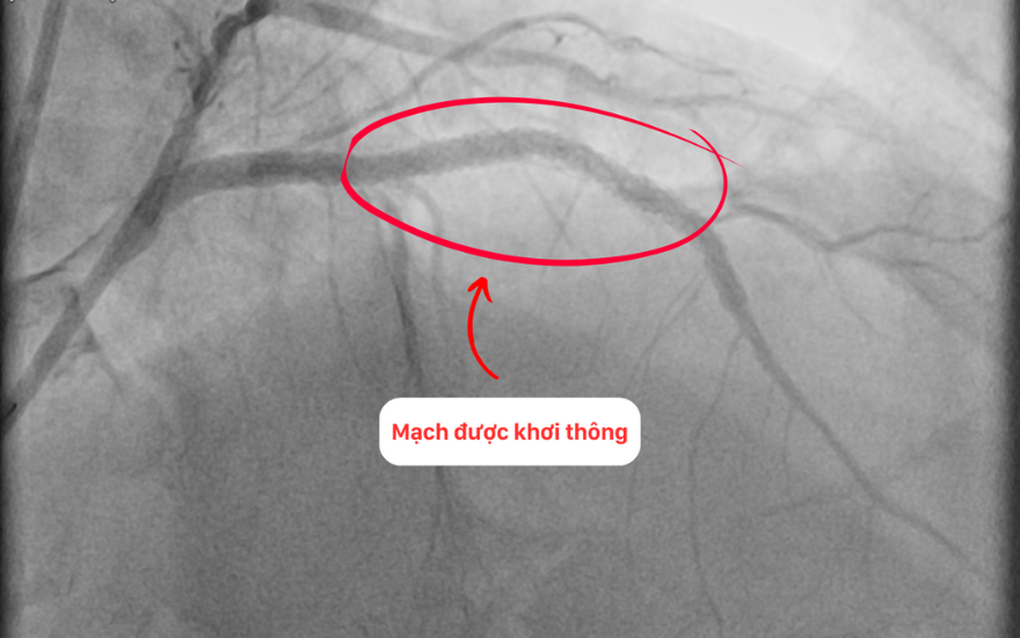
The circuit is re-opened.
Dr. Hiep said that using balloon cutting is an effective solution to treat atherosclerotic lesions, calcification, or restenosis in stents like Mr. Trong's case.
After the intervention, Mr. Trong felt relieved, comfortable, no longer had chest pain and could walk and move gently.
Dr. Hiep also warned that restenosis can develop silently. Therefore, patients should see a doctor early if they have any unusual symptoms such as chest pain, shortness of breath, fatigue, etc.
The doctor also emphasized that maintaining full medication as prescribed, regular check-ups, and changing daily habits, diet, and exercise as prescribed play an important role in long-term, safe, and effective management of coronary artery disease.
Source: https://dantri.com.vn/suc-khoe/can-thiep-thanh-cong-cho-benh-nhan-tai-hep-nang-mach-vanh-sau-15-nam-dat-stent-20250522185427391.htm



















Comment (0)