
Experts say that kidney stones are round or polygonal, usually 1 or more stones located in the lower or middle kidney.
Kidney stones are triangular or polyhedral in shape, usually 10-30mm in size, shaped like the kidney pelvis with the pointed end facing the spine. When the stone completely covers the renal pelvis, it is called coral stone, measuring 30-40mm.
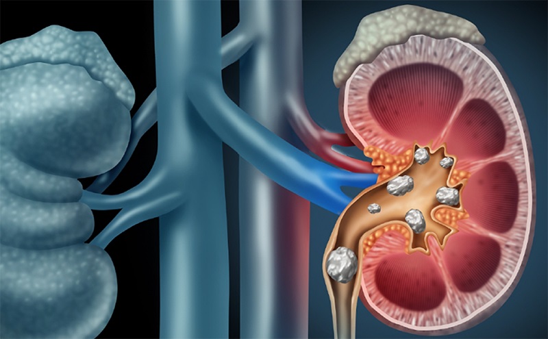
Kidney stones in many cases require surgery (photo source: Viet Duc Friendship Hospital).
Geographical, climatic and dietary factors influence the formation of kidney stones. Causes of stone formation include: Metabolic disorders causing hypercalcemia and hypercalciuria;
Changes in urine pH (normal pH: 5.6 - 6.3); congenital urinary tract malformations or acquired urinary tract stenosis causing urine retention create favorable conditions for stone formation.
Most cases of calcium stones have no known cause. Some cases of increased calcium are due to diet, diseases such as dehydration, prolonged immobility, hypercalciuria causing stones, or hyperparathyroidism causing increased calcium and decreased phosphorus.
According to doctors, the symptoms of this disease are dull pain in the lower back when kidney stones or coral stones have not caused obstruction. Sometimes patients have no symptoms and kidney stones are discovered during a routine health check-up or due to high blood pressure.
Renal colic is a typical pain when a stone blocks the renal pelvis and ureter. The pain radiates to the pelvis and scrotum, accompanied by vomiting and abdominal distension.
Blood in urine is caused by stones moving during movement or by infection causing damage to the renal pelvis mucosa and bleeding.
When there is a urinary tract infection: The patient has a high fever of 38o - 39oC, painful enlarged kidneys, cloudy urine and sometimes experiences septic shock with sweating, purple spots all over the body and low blood pressure.
Clinical examination shows enlarged and painful kidneys when kidney stones cause renal hydronephrosis. In some cases, patients come late for examination and find the lumbar region on the side with kidney stones swollen and red due to kidney stone obstruction causing renal pus retention, inflammation around the kidney or lumbar pus leakage due to perinephric abscess ruptured retroperitoneally and exposed to the skin.
When left late, renal pelvis stones will cause complications such as: Urinary tract infection; Pyelonephritis, interstitial nephritis, renal pelvis stenosis; Renal pelvis dilation, hydronephrosis, pyonephrosis, renal abscess; Fibrosing perinephritis (fibrose - xanthogranulomatosis); High blood pressure due to renal pelvis stones causing renal parenchymal ischemia, renal atrophy; Renal failure due to bilateral renal stones causing obstruction.
Regarding treatment, currently, percutaneous nephrolithotripsy (PCNL) is an interventional method using an endoscopic lithotripsy machine and removing kidney stones through a tunnel created through the skin, indicated for treating renal pelvis stones larger than 2cm with more benefits than open surgery.
Currently, percutaneous nephrolithotomy is the optimal and radical treatment method for kidney stones, replacing most of the indications for open surgery to remove stones. This method can crush large and solid stones. Direct percutaneous nephrolithotomy can wash away all the stone residue and drain the renal pelvis through the skin, allowing the treatment of most common kidney stones in our country.
Flexible ureterorenoscopy (FURS) is a method of using a flexible endoscope to pass retrogradely through the ureter to the renal pelvis to break up kidney stones using laser energy. The broken stones are removed by irrigation or using a Dormia or Nitinol basket to remove the stones.
This is an effective and safe treatment for kidney stones, indicated for kidney stones less than 2.5cm in size. Extracorporeal shock wave lithotripsy (ESWL): Indicated for the treatment of small renal pelvis stones with a diameter of £ 20mm, non-dilated renal pelvis, no infection, good renal morphology and function.
Kidney stones will break into small pieces less than 4mm in diameter and will be excreted through the urinary tract. Kidney stones measuring 20-30mm in size require a ureteral catheter to be placed before ESWL to prevent stone fragment blockage.
In addition to lithotripsy, many patients also have to undergo surgery. Renal pelvis stones have complications of severe recurrent hematuria, infection, pyelonephritis, hydronephrosis, pus retention, and renal failure. Complex renal coral stones with multiple stones have caused complications. Large bilateral renal stones will require surgery on the functional kidney first. In addition, there are many other treatment methods.
Source


![[Maritime News] More than 80% of global container shipping capacity is in the hands of MSC and major shipping alliances](https://vphoto.vietnam.vn/thumb/402x226/vietnam/resource/IMAGE/2025/7/16/6b4d586c984b4cbf8c5680352b9eaeb0)


Comment (0)