
Ho Chi Minh City: A 56-year-old man was rushed to the emergency room when one of his two thoracic aortic aneurysms ruptured, threatening his life. He was promptly saved by doctors at Gia Dinh People's Hospital.
On October 22, Dr. Tieu Chi Duc, Deputy Head of the Department of Thoracic and Vascular Surgery, said that the patient was admitted to the hospital with left back pain that radiated to the chest. Before that, the patient suddenly had back pain while riding a motorbike, the pain radiated to the chest and became worse when taking a deep breath.
Doctors assessed the patient in a very dangerous condition because the aneurysm in the aorta (the body's largest main artery) in the chest was likely to have ruptured. In addition, the condition of both lungs was very bad due to tuberculosis, with the risk of death at any time.
Doctors from many specialties urgently consulted to find a solution. CT scan recorded images of two thoracic aortic aneurysms, the first aneurysm measuring 63x75 mm had ruptured, the second aneurysm measuring 70x68 mm. Choosing a treatment method at this time was very difficult because the patient's lungs were very bad due to progressive tuberculosis, if surgery required anesthesia it would be almost impossible to succeed.
The team decided on the intervention plan through a stent graft catheter in the artery at the aneurysm location. This is a minimally invasive technique, reducing mortality and complications compared to open surgery.
The patient had a ruptured aortic aneurysm, but thanks to the surrounding structures such as the spine and lungs, the fragile localized structure was preserved, helping to preserve his life. Just a slight impact such as increased blood pressure or shaking the patient could cause the hematoma to rupture, threatening his life. Moreover, the patient's lungs were damaged, requiring the team to have experience with proficient techniques and a firm grasp of anatomy during the intervention.
"We almost 'held our breath' until we were able to pop the graft across the two ends of the blood vessel and isolate the ruptured aneurysm. Only then did we breathe a sigh of relief, knowing that we were able to save the patient's life," said Dr. Duc.
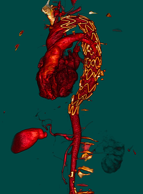
Image of blood vessels recovering circulation after intervention. Photo: Provided by the hospital
Post-operatively, the patient was closely monitored to prevent complications. A CT scan showed that the thoracic aorta at the site of the previous rupture had recovered stably. The patient was discharged after 5 days of treatment and transferred to his locality to continue taking tuberculosis medication.
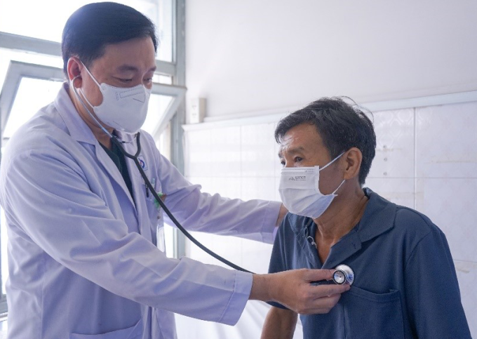
Doctor examines patient before discharge. Photo: Bich Hanh
The thoracic aorta originates from the heart, is the largest artery in the body, when ruptured causes acute cardiac compression, ischemia of organs such as the brain, liver, kidney... causing the patient to die quickly. Aortic aneurysm is a common disease today, occurring mainly in the elderly with comorbidities such as high blood pressure, diabetes, dyslipidemia, smoking habits.
Doctors recommend that patients with risk factors should go to medical facilities for early screening and timely treatment.
Le Phuong
Source link


![[Photo] General Secretary To Lam attends the 1st Congress of the Central Party Committee of the Fatherland Front and Central Mass Organizations](https://vphoto.vietnam.vn/thumb/1200x675/vietnam/resource/IMAGE/2025/9/23/2aa63d072cab4105a113d4fc0c68a839)


Comment (0)