


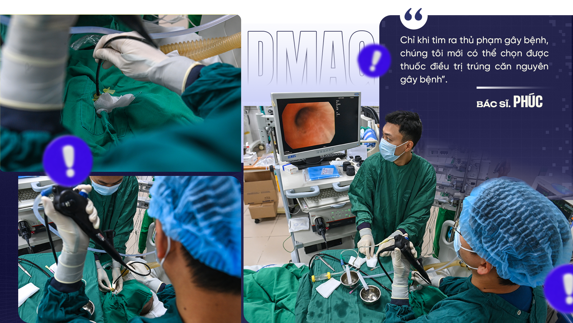
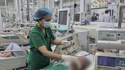
“Prepare the equipment, protective gear, and get to work,” said Dr. Pham Van Phuc, Deputy Director of the Intensive Care Unit at the National Hospital for Tropical Diseases. As soon as he finished speaking, the entire team busily moved around the intensive care unit. An emergency bronchoscopy procedure was immediately initiated.
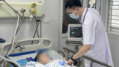
The 40-year-old woman lay motionless, her body emaciated after months in the hospital. She had undergone aortic arch replacement surgery at a central hospital, and was then transferred to the provincial hospital for monitoring.
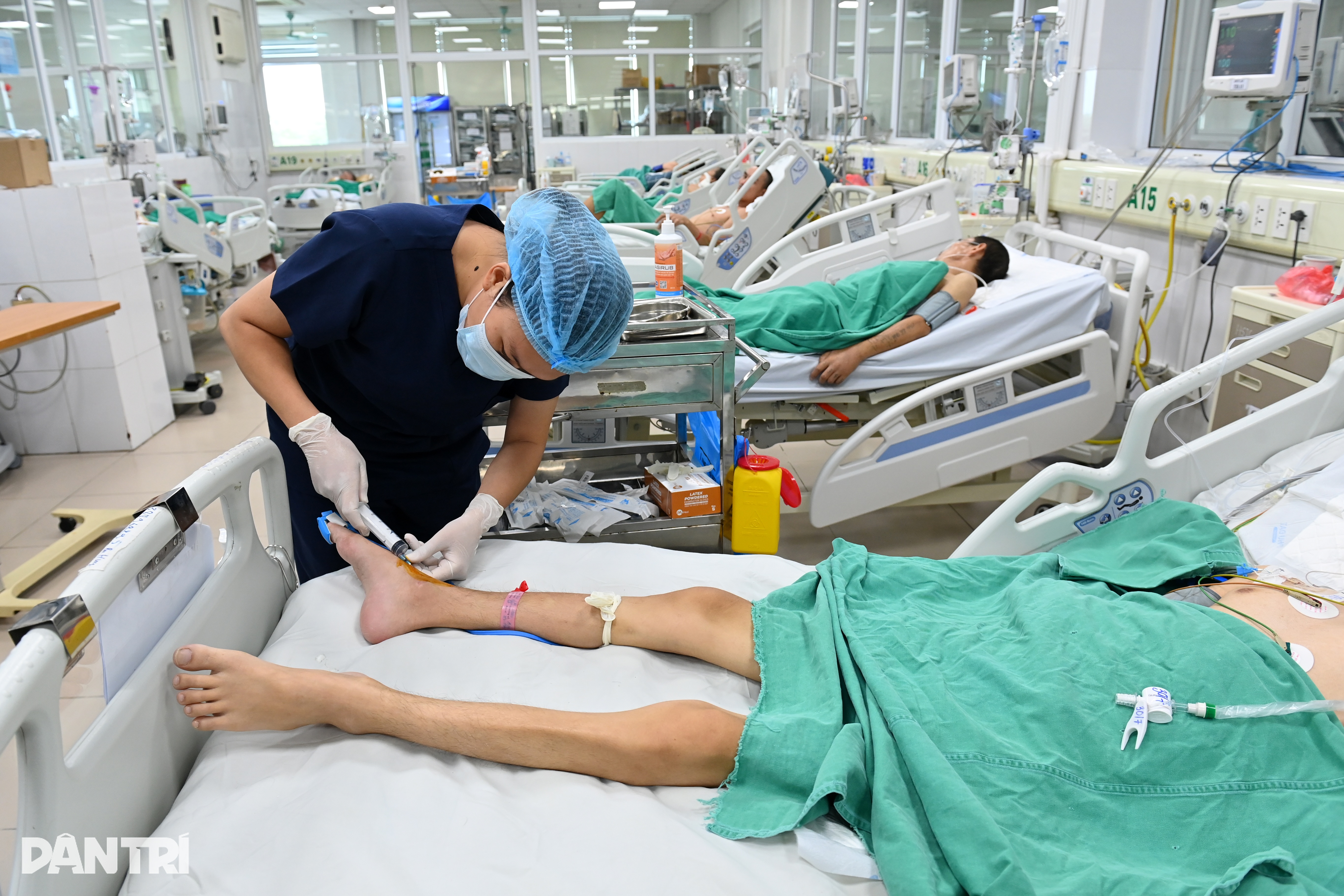
However, the long hospital stay allowed bacteria to "take over" her body like an invisible enemy.
At the provincial hospital, the patient was diagnosed with multidrug - resistant Pseudomonas aeruginosa infection.
This type of bacteria is resistant to most common antibiotics. After a month of treatment, the patient's condition did not improve. The high fever persisted, her breathing became increasingly rapid, and eventually she went into septic shock and had to be transferred to the National Hospital for Tropical Diseases.
The endoscope slid deep into the airway, revealing streaks of bright red, swollen mucous membrane on the screen.
Dr. Phuc explained: "The important goal is to obtain the deepest possible sample, at the exact site of infection, to determine the cause. Only when we find the culprit causing the disease can we choose a treatment that targets the root cause."

For patients dependent on ventilators, the risk of infection is ever-present. Pseudomonas aeruginosa, methicillin-resistant Staphylococcus aureus (MRSA), Klebsiella pneumoniae , and Acinetobacter baumannii are familiar but also haunting names for intensive care physicians.
They not only hide in the respiratory tract, but can also invade the bloodstream, brain and meninges, urinary and digestive systems, causing patients to rapidly develop multiple organ failure.
In such cases, microbiological testing and antibiotic susceptibility testing are the "lighting beam." They help identify which bacteria are present, which antibiotics are resistant or sensitive to them, and even whether those bacteria carry drug-resistant genes.
This is crucial for doctors to develop accurate treatment plans, instead of fumbling in the dark.
This 40-year-old patient is just one of dozens of cases of bacterial infection sampled every day. There are elderly women over 80 years old with recurrent hospital-acquired pneumonia, and healthy young men who suddenly collapse from encephalitis accompanied by infection.
The common thread is that they all need an answer: What is the real culprit? And what medications are still effective in saving them?

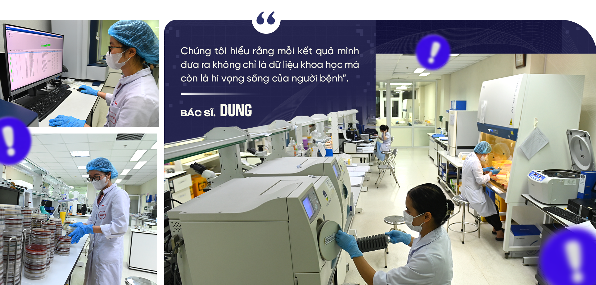
The destination for samples seeking analysis, the Department of Microbiology and Molecular Biology, with its state-of-the-art equipment and the bustling activity of its staff, can receive samples 24/7. It's considered a "tracing center" for pathogens.
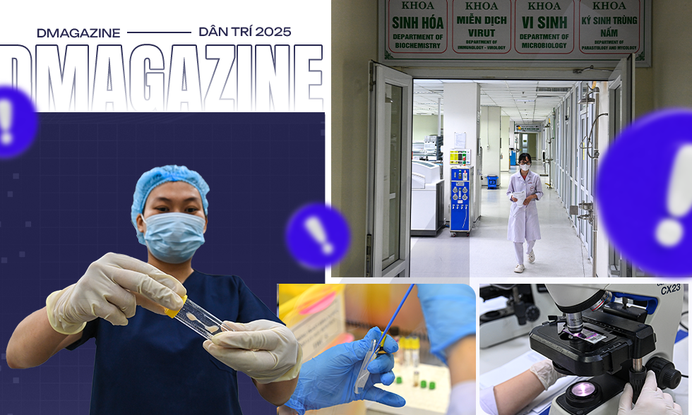
Each specimen from the clinical departments is treated as a valuable "clue." Upon receipt, technicians scan the code to display patient information, ensuring the sample is not misidentified. The data is immediately updated into the system, connecting it to the entire hospital.
In the sample transport box, the patient's blood and sputum samples had just arrived. Nurse Le Thi Thuy Dung quickly handed them over to her colleagues in the Microbiology laboratory. The blood samples were cultured to increase bacterial growth in a special medium, while the sputum samples had to be processed to remove impurities before culturing.
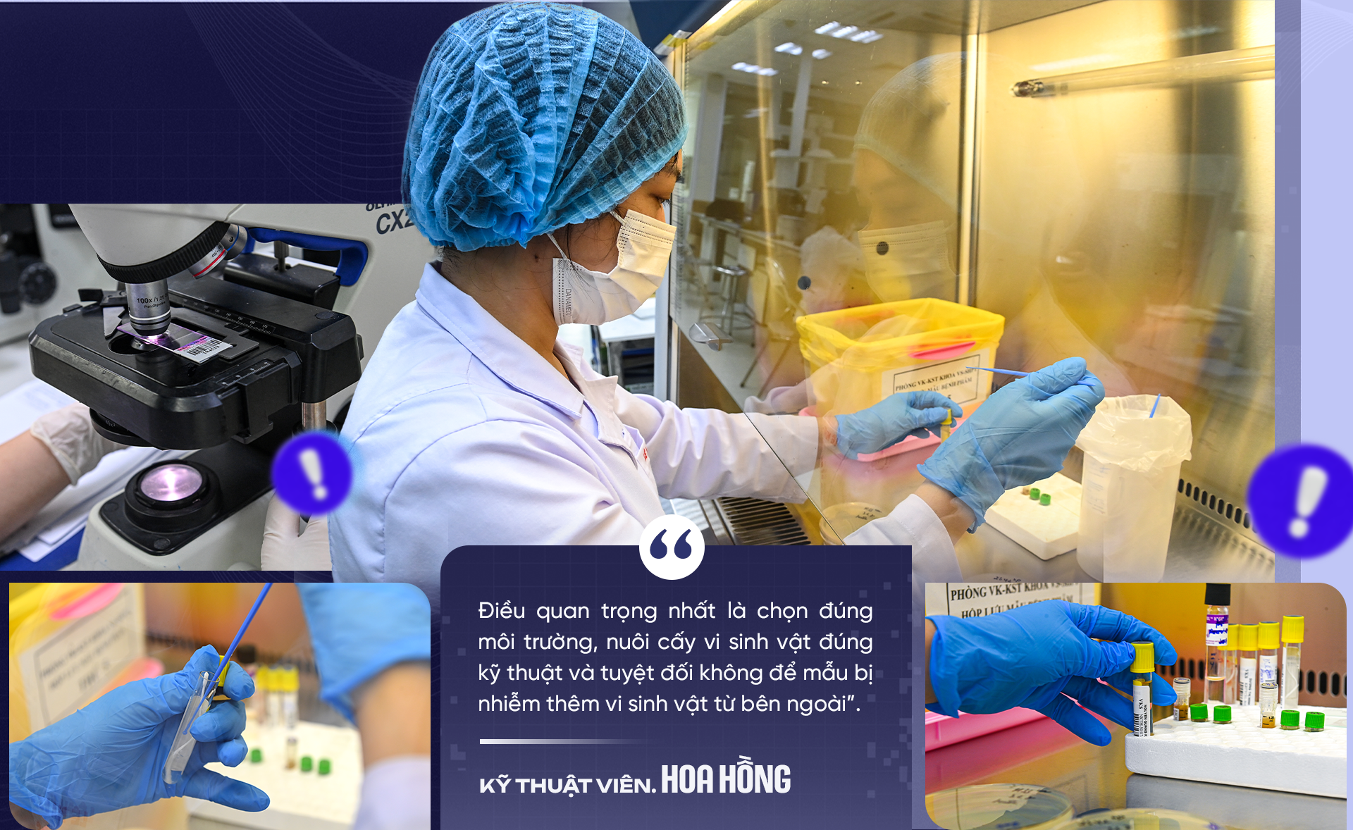
"The most important thing is to choose the right environment, cultivate microorganisms using the correct techniques, and absolutely prevent the sample from being contaminated with additional microorganisms from the outside," shared Le Thi Hoa Hong, a technician with many years of experience.
The procedure is performed in biosafety equipment, with each step of inoculating the specimen (which may contain pathogens) into the specific nutrient agar plate being carried out precisely. The inoculation loops are disposable and sterilized with gamma radiation before touching the sample.
The plates inoculated with bacteria are then placed in an incubator, where ideal temperature and humidity are maintained for their growth. This process lasts from 24 to 72 hours, or longer depending on the growth of each microorganism.
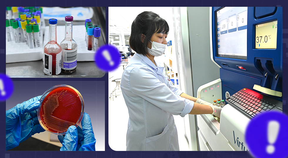
After the incubation period, tiny colonies begin to appear on the agar plate – traces of bacteria.
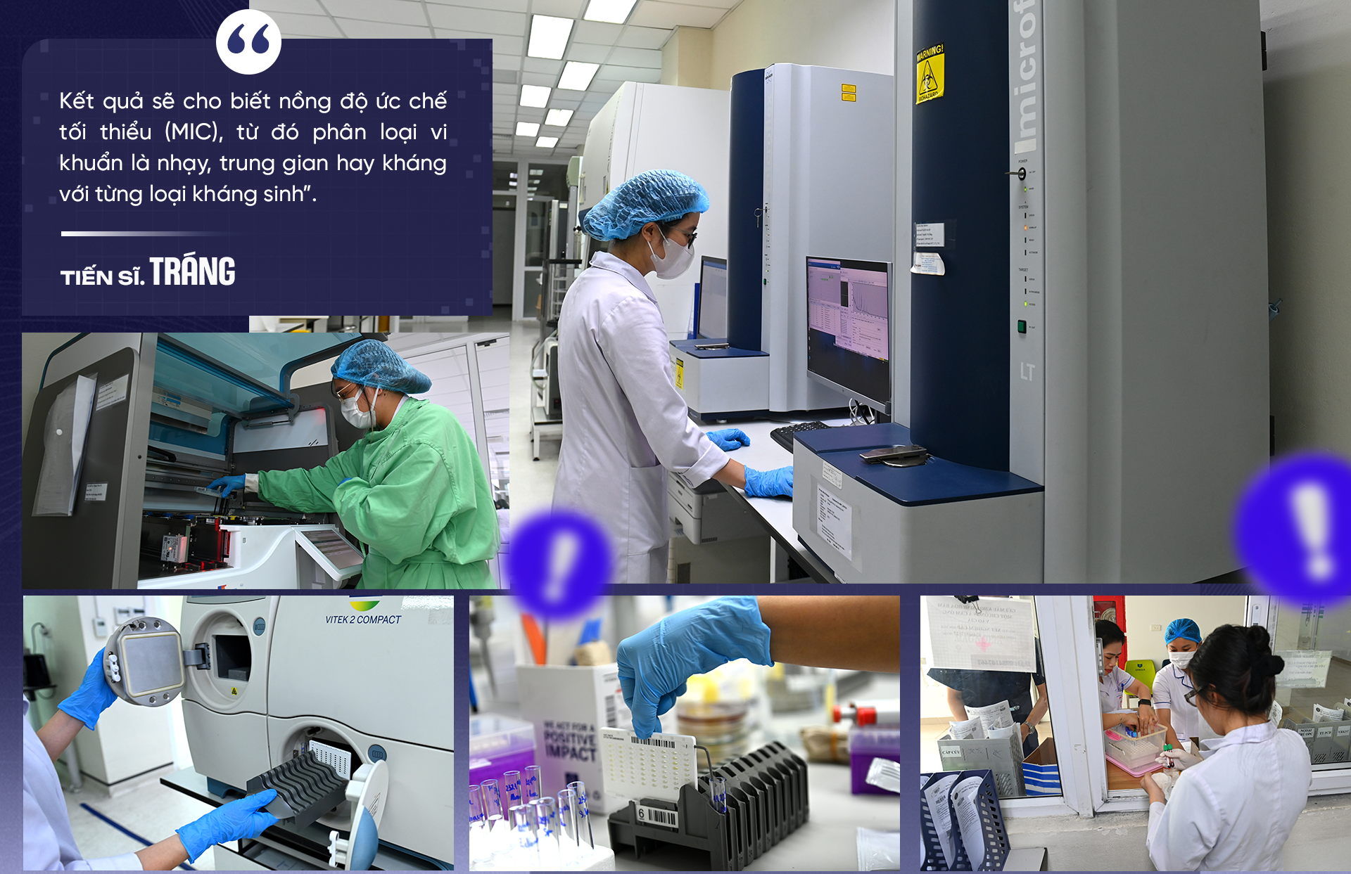
Technician Hong and her colleagues select suspected bacterial colonies, standardize turbidity, and then input them into identification and antibiotic susceptibility testing cards, before transferring them to the Vitek 2 compact automated system.
The machine will identify bacteria based on biochemical reactions and simultaneously perform an antibiotic sensitivity test, which involves "testing" the bacteria against a range of antibiotics to determine which drugs are still effective and which have become resistant.
"The results will show the minimum inhibitory concentration (MIC), thereby classifying the bacteria as sensitive, intermediate, or resistant to each antibiotic," shared Dr. Van Dinh Trang, Head of the Department of Microbiology and Molecular Biology.
However, the machine doesn't always have enough antibiotics available for testing.
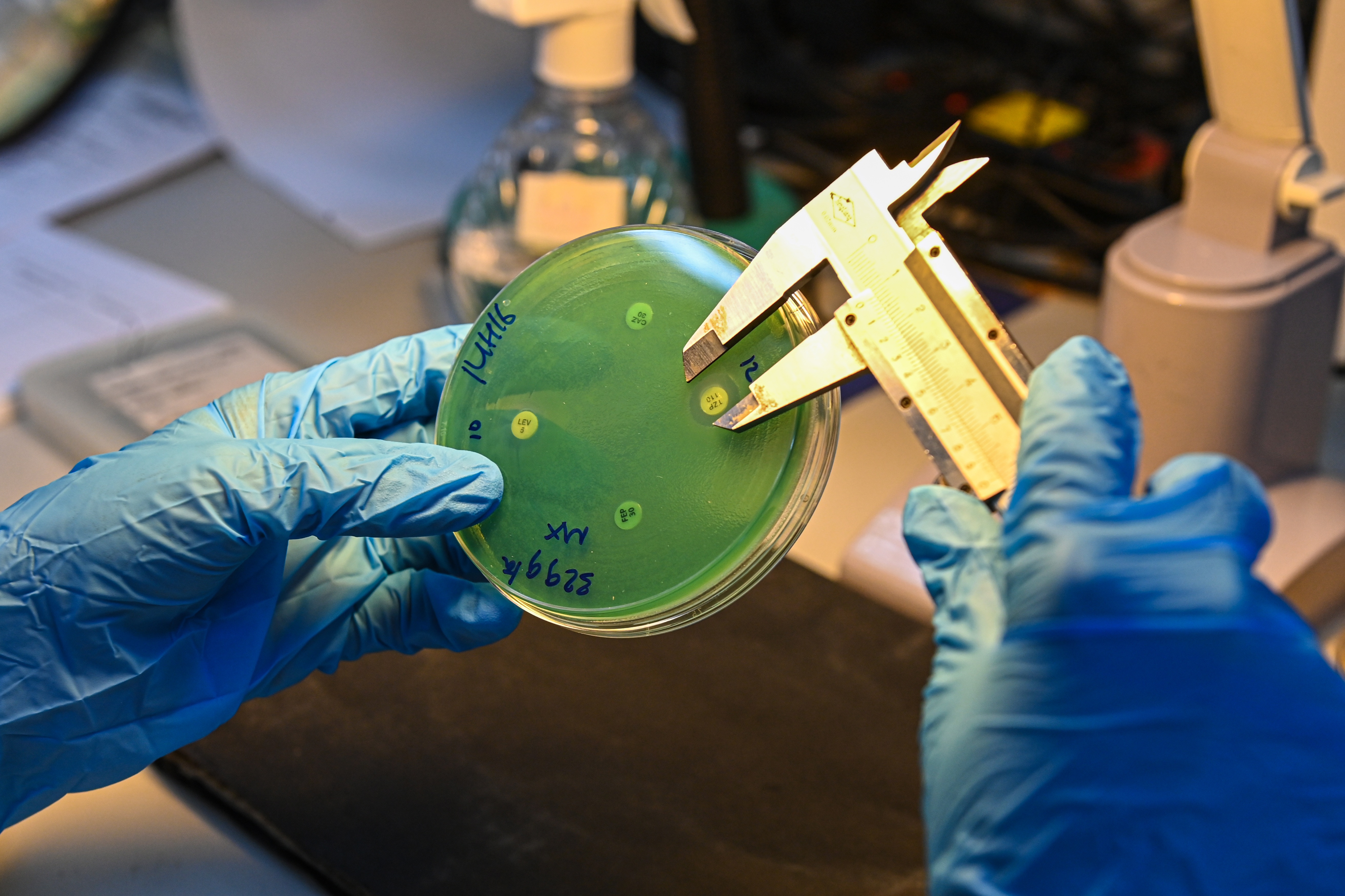
According to Dr. Trang, for rare or unusual bacterial strains exhibiting resistance, technicians must revert to the traditional method: using pre-soaked paper discs containing antibiotics at a specific concentration to diffuse the antibiotics into the agar plate.
On a petri dish, individual pieces of antibiotic-impregnated paper are placed on the surface of the agar plate inoculated with bacteria, and the diameter of the inhibition zone is measured to determine the level of antibiotic sensitivity or resistance of the bacteria.
Another helpful tool is the MALDI-TOF machine. This technology, which uses the characteristic protein spectrum of bacteria, can provide results in just a few minutes per sample.
"Each identification tray can hold up to 96 different samples. This allows us to process dozens of specimens in a single session, significantly reducing patient waiting times," explained Dr. Pham Thi Dung from the Department of Microbiology and Molecular Biology.

Even after the samples have been cultured and the microorganisms identified, the work of the Microbiology department staff doesn't stop there. This is when they enter the crucial stage: reading and analyzing antibiotic susceptibility testing.
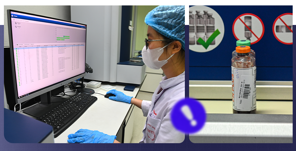
At her desk, Dr. Pham Thi Dung intently gazed at the screen displaying the results from the Vitek system. The data table was densely packed with symbols, and the MIC (minimum inhibitory concentration) index appeared next to the name of each antibiotic.
For each type of bacteria, the system automatically suggests a level of sensitivity, intermediate resistance, or resistance. However, before being forwarded to the clinician, all results must be confirmed by microbiology laboratory staff who will verify, cross-check, and approve them.
"The machine only provides raw data. Our job is to analyze whether the results are reasonable and consistent with the characteristics of this type of bacteria. If we find anything unusual, we have to do further testing using other methods," Dr. Dung shared.
Sometimes, a strain of bacteria exhibits resistance to most drugs in the antibiotic pool. In such cases, technicians must conduct additional genetic testing to determine if the bacteria carry any specific drug-resistant genes.
Only by knowing the specific "weapons" that bacteria possess can doctors choose the right medication to kill or counteract them.
During the peak of the Covid-19 pandemic, the workload at this "contact tracing center" increased many times over.
“There were days when we practically ate and slept right in the lab. The phone would ring with a new case, and everyone would immediately get into position, working through the night to get the results as quickly as possible,” Dr. Dung recalled.
Once the final results are available, the female doctor will prepare a detailed report, clearly stating the type of bacteria and its sensitivity to each antibiotic. "I always analyze the results using a tiered antibiotic system, identifying priority and contingency drug groups, so that clinicians have a basis for choosing the most optimal option," Dr. Dung explained.
A test result sheet may only contain a few lines of text, but behind it lie hours of meticulous, professional work. It can determine whether a patient's life is saved or not.
"We understand that every result we provide is not just scientific data, but also a glimmer of hope for patients," said Dr. Dung, her eyes still fixed on the antibiotic diffusion circles on the culture dish.

A week after receiving the antibiotic sensitivity test results from the Microbiology and Molecular Biology department, the 40-year-old woman was able to sit up on her own for the first time. Smiling, she thanked the doctors, saying, "I thought I had no chance."
That recovery began with the antibiotic sensitivity test results being sent to the intensive care unit. From the detailed data sheet on the type of bacteria and their sensitivity/resistance to each drug, the treating physician was able to develop a targeted treatment plan.
The highly drug-resistant Pseudomonas aeruginosa bacteria, which had previously caused patients to go into shock and experience persistent high fever, were eventually brought under control. Respiratory indicators stabilized, and the fever gradually subsided.
On the day she was discharged from the hospital, the whole family embraced each other at the hospital gate. This joyful reunion was made possible by the silent but crucial contribution of the "bacteria hunters." They weren't present at the bedside, didn't hold stethoscopes or needles, but every result they returned played a decisive role in giving patients a chance at survival.
Source: https://dantri.com.vn/suc-khoe/ven-man-nghe-la-cua-nhung-tho-san-vi-khuan-20251014160424246.htm
![[Image] Pleasant weather helps students enter the 10th grade entrance exam with confidence.](https://vphoto.vietnam.vn/thumb/1200x675/vietnam/resource/IMAGE/2026/05/29/1780034401612_ngay-1-thi-lop-10-minh-duy-8-5009-jpg.webp)




![[Photo] Ba Lang An Lighthouse - the "eye of the sea" amidst the "rock museum" of Quang Ngai province.](https://vphoto.vietnam.vn/thumb/1200x675/vietnam/resource/IMAGE/2026/05/29/1780038698840_anh-man-hinh-2026-05-29-luc-14-10-42.png)






































































Comment (0)