
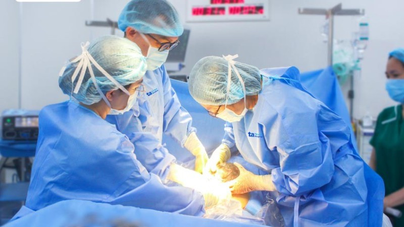
The surgeon actively performed surgery on the pregnant woman.
The twins are the result of in vitro fertilization of Ms. D and her husband after 6 years of infertility. At week 12, an ultrasound at the Fetal Medicine Center, Tam Anh General Hospital, Ho Chi Minh City, recorded a fetus with a 3.5mm thick nuchal translucency and subchorionic fluid accumulation.
Specialist Doctor I Le Quang Hung, Center for Fetal Medicine, explains that a nuchal translucency of 3.5mm is a high-risk sign indicating that the fetus may have chromosomal abnormalities (Down, Patau, Edwards syndromes...), gene mutations or congenital heart defects. Subchorionic hematoma is a condition in which blood accumulates between the placenta and the uterus, which can threaten miscarriage.
The doctor advised invasive testing (chorionic villus sampling or amniocentesis) for both fetuses to check for genetic abnormalities, but Ms. D and her husband refused. The pregnant woman was then monitored through each stage of her pregnancy, paying close attention to examining the morphology of the fetus, especially congenital heart abnormalities.
The pregnant woman was in the high-risk preeclampsia group and was given aspirin to prevent preeclampsia. Dr. Hung instructed her to eat a healthy diet, limit sweets and starches, increase protein, green vegetables, unsweetened fresh milk, divide meals into smaller portions, do light exercise, and check blood sugar and blood pressure at home.
At 29 weeks of pregnancy, Doppler ultrasound recorded 2 fetuses with intrauterine growth restriction, risk of fetal heart failure, stillbirth, premature birth, respiratory failure after birth... The doctor monitored every week to assess the health indicators of the fetus.
At week 32, the smaller fetus has increased umbilical artery PI (reduced ability to supply oxygen and nutrients through the placenta) and decreased CPR (reduced blood flow to other organs to prioritize blood and oxygen to the brain). Therefore, the mother has a Doppler ultrasound every 3 days. By week 37, the amniotic fluid index of the smaller fetus has decreased to 1.8cm (normal is 2-8cm for twins).
At this time, if the fetus is growth-restricted with oligohydramnios in the uterus, the risk is higher than premature birth. Therefore, Dr. Hung ordered an active cesarean section for the mother at week 37 to ensure the safety of both babies. The twins were born safely, one weighing 2.5kg, the other about 2.3kg, and in stable health.
According to Dr. Hung, the cause of intrauterine growth restriction can come from the mother (high blood pressure, chronic diseases, etc.), from the fetus (chromosomal abnormalities, malformations, fetal infections, etc.) or from the placenta (impaired placental function).
Accurate assessment of gestational age is the condition to determine whether the size of the fetus corresponds to the gestational age. According to the International Society of Ultrasound in Obstetrics and Gynecology (ISUOG), measuring the fetal crown-rump length in the first trimester (11-13 weeks and 6 days) is the optimal method to calculate gestational age.
Doppler ultrasound helps assess blood flow in the umbilical artery - the blood vessel that supplies oxygen and nutrients to the fetus, contributing to determining whether the fetus has intrauterine growth restriction.
There is currently no way to prevent this condition. Doctors recommend that pregnant women comply with regular prenatal checkups. Early diagnosis, monitoring and evaluation of fetal health, and choosing the appropriate time to end the pregnancy can reduce the mortality rate or risk of disease for the child later.
HAI NGO
Source: https://nhandan.vn/duong-them-2-thang-cho-san-phu-mang-song-thai-co-gioi-han-tang-truong-trong-tu-cung-post889465.html


Comment (0)