

According to information from Bai Chay Hospital ( Quang Ninh ), doctors in the Respiratory Internal Medicine Department successfully removed a foreign object from the apical lobe of the left lung of an elderly patient using flexible bronchoscopy.
The patient, Mr. NVT (73 years old, residing in Quang Ha commune, Quang Ninh province), had a history of smoking for many years at a rate of approximately 30 packs/year and regularly consumed alcohol. He was admitted to the hospital with chest pain, a persistent cough for more than a month, expectoration of thick phlegm, chest pain radiating to the back, but no fever.
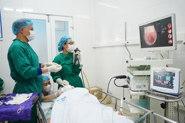
The intervention team from Bai Chay Hospital performed flexible bronchoscopy to investigate and determine the cause of the illness.
Previously, the patient had been treated at a lower-level hospital with a diagnosis of pneumonia and suspected lung tumor, but their condition did not improve, so they were transferred to Bai Chay Hospital.
At the hospital, a chest CT scan revealed consolidation in the left lung, thickening of the interstitial tissue in both lungs, and dilation of some bronchial branches in the lower lobe of the left lung. These lesions could easily be mistaken for serious respiratory diseases such as lung tumors or tuberculosis.
Following a consultation, the intervention team, led by Dr. Pham Thi Ut Trang, Deputy Head of the Department of Respiratory Medicine at Bai Chay Hospital, performed flexible bronchoscopy to investigate and determine the cause.
During the endoscopy, doctors discovered a foreign object lodged deep in the apical bronchus of the left lung, accompanied by mucosal hyperplasia, edema, and a high risk of bleeding. This is a rare location for an airway foreign body; the bronchial lumen is small, the angle of access is difficult, and there is a risk of mucosal damage or pushing the foreign object deeper.
Using skillful techniques and experience in handling complex airway foreign body cases, the team successfully removed the foreign body from the bronchus, relieving the airway obstruction. Following the intervention, the patient's symptoms of coughing and chest pain improved significantly.
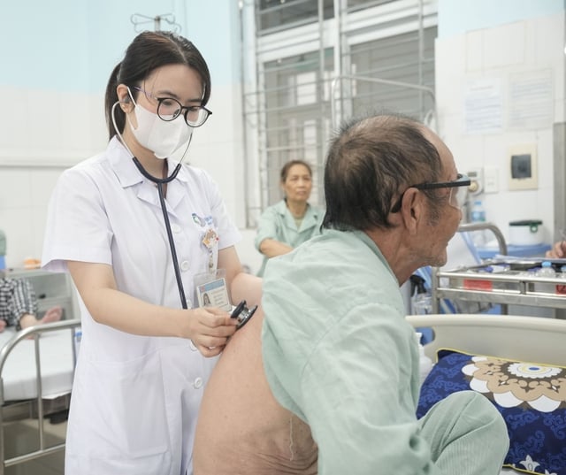
The patient's health is stable after the intervention.
According to Dr. Pham Thi Ut Trang, airway foreign bodies are often difficult to detect due to atypical symptoms, easily mistaken for chronic pneumonia, pulmonary tuberculosis, or lung tumors, especially in older adults, those with underlying respiratory conditions, or a long history of smoking.
According to Dr. Trang, if not detected early, patients may face dangerous complications such as recurrent pneumonia, lung abscess, lung collapse, or chronic respiratory dysfunction. For foreign bodies located deep within the segmental bronchi, intervention requires an experienced endoscopist with good control of the endoscope and precise manipulation to avoid pushing the foreign body deeper or damaging the airway.
Therefore, doctors advise people that if they experience persistent coughing, chest pain, phlegm production, and pneumonia that does not improve after several days of treatment, they should go to a specialized medical facility for timely examination and diagnosis to avoid overlooking the cause, which may be a foreign object in the airway.
Source: https://suckhoedoisong.vn/gap-thanh-cong-di-vat-phe-quan-o-vi-tri-hiem-gap-169260527140329249.htm
![[Photo] Standing Committee member of the Party Central Committee Tran Cam Tu working with the Central Inspection Committee](https://vphoto.vietnam.vn/thumb/1200x675/vietnam/resource/IMAGE/2026/05/28/1779969579668_ndo_br_bnd-2495-jpg.webp)


























































































Comment (0)