On April 16th, Dr. Nguyen Anh Dung, Head of the Cardiovascular and Thoracic Surgery Department at the Cardiovascular Center, Tam Anh General Hospital, Ho Chi Minh City, stated that the CT scan revealed a 27x25 mm lung tumor located near the left hilum of the patient. Most lung tumors or nodules are asymptomatic in their early stages. In Ms. H.'s case, the tumor was located near the hilum of the lung, close to the heart, causing her to experience chest tightness, although not severe. She sought medical attention and the tumor was detected early.
The team performed minimally invasive procedures for preliminary biopsies on the patient, including bronchoscopy and CT-guided lung tumor biopsy, but both yielded benign results. However, based on experience and the imaging characteristics of the tumor, the doctors strongly suspected it was a non-benign tumor and decided that the entire tumor needed to be removed for diagnosis and treatment.
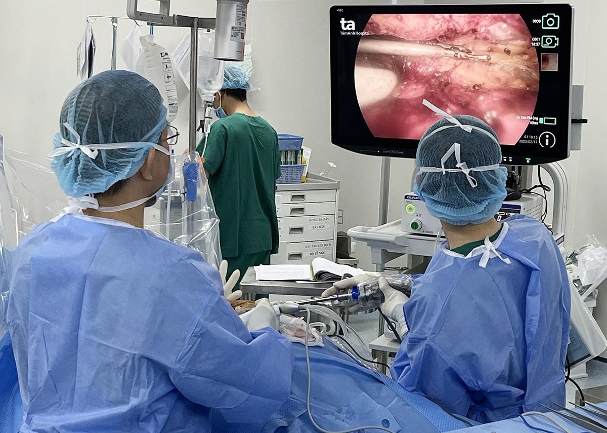
The patient underwent thoracoscopic tumor resection and frozen section biopsy. The surgeon extracted a tumor sample via the endoscope, which was then sent for pathological examination and frozen section biopsy in 30-45 minutes. The results confirmed it to be a malignant lung tumor.

Doctors in a laparoscopic surgery to remove a lung tumor.
According to Dr. Dung, endoscopic lung tumor biopsy or CT-guided lung tumor biopsy are minimally invasive methods that help diagnose lung lesions such as tumors and inflammation. Sometimes, the biopsy yields a false-negative result, meaning the tumor appears benign when it is actually malignant. This is because the tumor is located in a difficult-to-access area, and the biopsy did not hit the location of the malignant cells. In these cases, if the nature of the tumor is still suspected, the doctor will order endoscopic surgery to remove the entire tumor for frozen section biopsy.
Following the malignant test results from the surgery, Dr. Dung and his team decided to remove the upper lobe of the left lung along with a complete mediastinal lymph node dissection for Ms. H. in order to completely remove the tumor from her body and reduce the risk of cancer recurrence. The patient underwent only one surgery to address two issues: diagnostic biopsy and surgical treatment.
After the surgery, Ms. H. no longer experienced chest discomfort, and the small endoscopic incision resulted in minimal pain. She stated that she had not been exposed to cigarette smoke or harmful chemicals and had no family history of lung cancer. Subsequent genetic testing revealed an EGFR gene mutation. The patient was treated with targeted therapy according to a protocol to prevent cancer recurrence.
Source link


Comment (0)