
I am 28 years old and have a fibroadenoma in my left breast. Is this a form of breast cancer, dangerous? (Ngoc Chau, Ho Chi Minh City)
Reply:
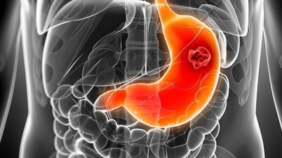
Fibroadenoma, also known as fibroadenoma, is a solid, round mass made up of fibrous and glandular tissue, usually benign. The disease is common in women aged 15-35, and is rarely seen in postmenopausal women.
Most fibroadenomas do not increase the risk of breast cancer. But they cause changes in the breast that make it harder to detect and diagnose cancer during a breast exam or mammogram.
In some cases, fibroadenoma increases the risk of cancer if the fibroadenoma is complicated or if there is a phyllodes tumor (leaf-like shape) associated with it. Small, uncomplicated fibroadenomas that grow slowly and increase in size with hormonal changes due to pregnancy do not require treatment. This type of fibroadenoma can increase in size and bleed internally, but this rarely occurs.

Most fibroadenomas are benign and do not increase the risk of breast cancer. Photo: Freepik
Tumors can form painless, easily movable lumps on one or both sides of the breast under the skin. Tumors can be as small as a pea or as large as 2-3 cm.
Typical fibroadenomas are round or oval in shape with a smooth, well-defined border, and tend to soften a few days before menstruation. Larger fibroids are more likely to cause pain. They tend to grow very slowly, and in many cases shrink, and may grow larger during pregnancy.
The disease is detected during a health check or during a routine mammogram or ultrasound. Some diagnostic methods are as follows:
Breast ultrasound : This method is used to evaluate breast tumors. On ultrasound, fibroadenomas are easily distinguished from other tissues due to their response to sound waves. They appear as dark areas with clear borders, similar to transparent, round or oval, with smooth concave and convex areas.
Mammography: Tumors appear as round or oval masses with clear borders that do not invade surrounding tissue. Sometimes they are accompanied by large calcifications (calcium deposits).
3D Mammography : A specialized mammography method that creates 3D images of the breast, providing more detail than a regular mammogram.
Biopsy : Doctors order a tissue sample to be taken from a person with a fibroadenoma for pathological examination. Different types of biopsies exist, depending on the purpose of diagnosis or treatment, including core needle biopsy, which uses a needle to take a tissue sample, and fine needle aspiration.
You did not indicate the size of your fibroadenoma. However, most fibroadenomas are small, slow-growing, stable, and do not require treatment. Some fibroadenomas shrink or disappear on their own. If the biopsy confirms that the tumor is not cancerous, the patient should return for a follow-up visit in 3-6 months so the doctor can monitor the tumor for any changes.
Master, Doctor Nguyen Do Thuy Giang
Head of Breast Surgery Department, Tam Anh General Hospital, Ho Chi Minh City
| Readers ask cancer questions here for doctors to answer |
Source link


![[Photo] Closing of the 14th Conference of the 13th Party Central Committee](https://vphoto.vietnam.vn/thumb/1200x675/vietnam/resource/IMAGE/2025/11/06/1762404919012_a1-bnd-5975-5183-jpg.webp)


Comment (0)