
Ms. H. (37 years old, in Ho Chi Minh City) had surgery to treat endometriosis and uterine fibroids, which severely reduced her ability to conceive naturally, requiring assisted reproduction with a small number of embryos. During her first pregnancy, the fetus had Edwards syndrome, so she had to terminate the pregnancy at week 19. After many efforts, she became pregnant for the second time. However, at week 17, a morphological ultrasound at the Center for Fetal Medicine, Tam Anh General Hospital, Ho Chi Minh City showed that the fetus had a 3-4 cm long sacral teratoma.
Despite the 17% risk of malignancy, the family decided to keep the pregnancy. At week 22, MRI showed the tumor growing rapidly, with many blood vessels, protruding from the body. By week 30, the tumor had grown five times larger, threatening the fetus.
Fetus with sacral teratoma
The doctor diagnosed the fetus with a type 1 sacral teratoma, the tumor was completely outside the body, with many blood vessels proliferating. Blood from the fetus poured into the tumor, causing the baby to gradually become anemic and have heart failure. By week 34, the tumor was twice as large as the fetus's body, the baby began to have heart failure, and the risk of the tumor rupturing caused hemorrhagic shock, threatening the lives of both mother and child.
The Obstetrics, Neonatology and Pediatrics departments plan to coordinate monitoring, cesarean section, care and surgery when the baby is healthy enough. Two adjacent operating rooms are prepared, one for the cesarean section and primary care team, the other for the tumor removal surgery team.
Fetus with sacral teratoma weighing 1.8 kg
PHOTO: D.L
Prolonged time, the fetus can die suddenly, the mother is at risk of postpartum hemorrhage
On May 19, Dr. Nguyen Ba My Nhi, Director of the Obstetrics and Gynecology Center, Tam Anh General Hospital, Ho Chi Minh City, said that in the case of pregnant woman H., if the situation continues, the fetus will die suddenly. If the tumor ruptures in the uterus, it will cause a lot of blood loss and threaten the lives of both mother and child.
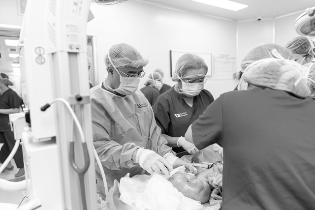
The doctors decided to make a vertical incision from the lower part to the bottom of the uterus, carefully removing the fetus safely without rupturing the tumor. Ms. H. had her uterine muscles quickly restored, no postpartum hemorrhage occurred and no blood transfusion was needed.
The baby was born safely, weighing 3.4 kg, with a tumor weighing 1.8 kg and nearly 20 cm long. At that time, the neonatal team gave the baby oxygen, provided postnatal care, and stabilized vital signs. The doctor decided to take the baby to the intensive care unit, and it is expected that after 24 hours, when the baby is stable, surgery will be performed to remove the tumor.
However, 2 hours later, bleeding began inside the tumor, the tumor grew larger, and the child was at risk of hemorrhagic shock, threatening his life. The hospital immediately activated the emergency mode for the entire hospital, immediately transferring the child to the operating room to perform surgery to stop the bleeding and remove the tumor.
Prevention of the risk of heavy blood loss during surgery
Specialist Doctor 2 Nguyen Do Trong said that the tumor is attached to the body, if separated, the child will lose a large amount of blood, so it is necessary to both resuscitate and perform surgery. At the same time, the patient is stored in the blood bank, preparing all blood products such as red blood cells, plasma, platelets, etc. to compensate for the child during and after surgery.
"The tumor is very large and may involve the colon, bladder, genitourinary organs and surrounding structures," said Dr. Trong.
After nearly 4 hours of surgery, the tumor was successfully removed and the patient was actively monitored. 24 hours after surgery, the health of both mother and child was stable, the mother ate and walked normally, the uterus contracted well, and there was no vaginal bleeding. The baby was taken off the ventilator after one day, circulation and respiration were stable, and the surgical wound was dry. The baby is now healthy and has been discharged from the hospital.
According to pediatricians, sacral teratoma is a rare disease (1/20,000-40,000 cases), including 4 types and unknown causes. The disease can be detected early through modern prenatal ultrasound, helping to plan treatment immediately after birth. Pregnant women need to have regular check-ups at a specialized facility to promptly detect congenital malformations.
Source: https://thanhnien.vn/hiem-gap-thai-nhi-mang-khoi-u-quai-to-gap-doi-co-the-185250519144929063.htm


![[Maritime News] More than 80% of global container shipping capacity is in the hands of MSC and major shipping alliances](https://vphoto.vietnam.vn/thumb/402x226/vietnam/resource/IMAGE/2025/7/16/6b4d586c984b4cbf8c5680352b9eaeb0)


Comment (0)