
The patient came to the hospital for a routine health check-up as required by the unit, but had registered for an additional cardiovascular examination due to occasional palpitations and left chest pain, and was suddenly found to have an abnormal mass in the lung.
Medical news on January 12: Health check-up detects rare lung defect
The patient came to the hospital for a routine health check-up as required by the unit, but had registered for an additional cardiovascular examination due to occasional palpitations and left chest pain, and was suddenly found to have an abnormal mass in the lung.
Health check-up detects rare lung defects, cancer risk
Recently, Medlatec General Hospital received Mr. VVĐ (45 years old, bank employee) for a routine health check-up as required by the unit and was unexpectedly diagnosed with isolated lung disease in the lower lobe of the left lung.
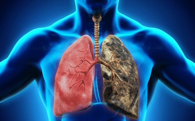 |
| Pulmonary sequestration is a rare congenital abnormality, accounting for only 0.15 - 6.4% of cases. Illustrative photo. |
Medical history revealed that Mr. D.'s father had lung cancer and he himself smoked. Recently, due to pain in the left chest and palpitations, Mr. D. registered for additional cardiovascular examination in addition to the items included in the unit's test package.
Chest X-ray results showed a opacity in the lower left lung area. Chest CT scan without contrast injection showed a solid mass in the lower left lobe of the lung, with irregular edges and clear boundaries, measuring 21x28x37mm, without air-filled bronchi, accompanied by a 7mm enlarged lymph node, leading to monitoring for a tumor in the lower left lobe of the lung.
For differential diagnosis, the patient was ordered a chest-abdominal CT scan with contrast injection. The results showed that the S6 segment of the left lower lung had a solid nodule that strongly enhanced the contrast after injection, with an abnormal blood vessel branching from the aorta to the feeding artery with a diameter of 5mm.
Based on the results of the imaging diagnosis, the doctor diagnosed the patient with isolated lung in the lower lobe of the left lung. Mr. D. was advised to have early surgery and regular follow-up every 3-6 months.
According to Dr. Tran Thi Thu, Department of Diagnostic Imaging, Medlatec Healthcare System, isolated lung is a rare congenital abnormality, accounting for only 0.15 - 6.4%.
This is a condition of dysplastic lung parenchyma, supplied by systemic arteries without communication with the tracheobronchial tree, leading to changes in respiratory structure and function.
Lobar sequestration (75-85%): Often seen in older children or adults, with typical symptoms of acute pneumonia or recurrent pneumonia. Most of these lesions are found next to the diaphragm or the posterior basal segment of the left lower lobe, and the draining vein will drain into the pulmonary vein.
Extralobar sequestration (15 - 25%): Common in newborns, with signs such as respiratory failure, cyanosis or infection. Venous drainage flows into the systemic veins and right atrium.
The disease forms when an accessory lung bud develops lower than the normal lung buds and receives its blood supply from the primitive foregut vessels.
If the accessory lung bud develops before the pleura forms, both the normal lung tissue and the isolated lung tissue will be surrounded by the same pleura, resulting in an intralobar isolated lung. Conversely, the isolated lung tissue has its own pleura, resulting in an extralobar isolated lung.
Doctor Thu warned that if left untreated, isolated lung can lead to serious complications such as local inflammation, coughing up blood, recurrent infections and the risk of malignancy, becoming lung cancer.
In pulmonary sequestration, surgery is the mainstay of treatment, helping to prevent recurrent infection, hemoptysis, and the risk of malignancy. Interventions such as embolization may also be considered.
Because the clinical symptoms of the disease are often vague, regular health check-ups at reputable medical facilities with a team of highly specialized doctors are extremely necessary to detect the disease early, thereby intervening promptly, increasing the chance of successful treatment and minimizing complications.
Things to know about congenital pulmonary cystic anomalies in fetuses
Ms. Lanh, 33 years old, during her 23-week ultrasound, discovered a bright mass in the left lung of the fetus, measuring 45x30x32mm, containing many small cysts from 1-6mm inside.
According to doctors, this mass displaced the fetal heart to the right and had blood supply from the pulmonary artery. However, there were no signs of fluid in the body cavities and the amniotic fluid was normal. Doctors diagnosed the fetus with cystic adenomatous hyperplasia in the left lung, but there were no complications of fetal edema or heart failure.
Cystic adenomatous pulmonary hyperplasia (also known as congenital adenomatous pulmonary malformation) is a condition in which abnormal lung tissue forms during the embryonic stage. This tissue does not function like a normal lung, usually appears in only one lobe of the lung, and can develop into cystic or solid masses.
Currently, Ms. Lanh's pregnancy is 28 weeks, the size of the cyst has not changed and other indicators are stable. Doctor Nghi predicts that the situation will be good, no need for medication or special treatment, just close monitoring until birth.
After birth, the doctor will perform a chest CT scan to assess the size of the lesion and determine the time for surgical removal to avoid the risk of recurrent lung infection and prevent the possibility of progression to lung cancer later.
Ms. Vy, 28 years old, was pregnant for the first time. During the 18-week ultrasound, a luminal mass measuring 25x15x26mm was also detected in the left lung.
However, this mass did not displace the heart and there were no signs of fetal edema or fetal distress. Over the following weeks, the mass increased slightly in size and then decreased to 14x8x13mm.
By week 37, the ultrasound no longer detected the hyperfluorescent mass, and the baby was delivered vaginally at week 39. The baby was born healthy, breathing well, and at three months, a chest CT scan showed completely normal lungs.
Doctor Nguyen Thi Mong Nghi, Center for Fetal Medicine, Tam Anh General Hospital, Ho Chi Minh City, said that congenital pulmonary cysts change in size throughout pregnancy and are often detected by ultrasound with the manifestation of a bright mass that may or may not contain a cyst inside in the chest area of the fetus.
The mass may remain stable in size, grow in parallel with fetal growth, or even decrease or disappear during the third trimester.
Most cases have good outcomes. However, about 10% of tumors grow too large, threatening the life of the fetus. When the tumor is too large, it can cause pulmonary hypoplasia (underdeveloped lungs), compress blood vessels, increasing pressure on the heart, leading to heart failure, fetal edema, or prevent the fetus from swallowing amniotic fluid, causing polyhydramnios.
Congenital adenomatous pulmonary malformations are divided into five types, ranging from type 0 to type 4. Type 0 is very rare and usually causes death after birth, while type 4 has a risk of progressing to lung cancer later in life.
The cause of the disease is unknown, and it is not related to race, maternal age, genetic or chromosomal abnormalities, nor is it hereditary (except in some cases of type 4 associated with familial pleomorphic myeloproliferative neoplasia syndrome).
According to Dr. Nghi, fetuses with pulmonary cystic malformations need to be closely monitored via prenatal ultrasound to determine the type, size, location, and blood supply to the tumor, as well as assess other organ abnormalities.
In complex cases, the use of MRI can help assess the condition of the fetus more clearly. Depending on the specific condition of the fetus and the pregnant woman, the doctor may decide to intervene during pregnancy or closely monitor until birth and intervene after birth.
If the cyst is small and does not affect the heart or cause fetal hydrops, the doctor will usually continue to monitor the baby after birth. In most cases, the baby will not have breathing problems and the cyst can be surgically removed within 6-9 months after birth.
On the contrary, if the cyst grows rapidly and large, usually occurring between weeks 20-28 of pregnancy, immediate treatment is required to avoid risks such as fetal edema, heart failure, or death.
Doctor Nghi recommends that pregnant mothers should have regular check-ups to detect pulmonary cysts early, helping doctors closely monitor the health of the fetus and pregnant woman, thereby providing timely treatment plans to protect the health of both mother and baby.
Girl with epilepsy due to cerebral vascular malformation
According to information from the Xanh Pon General Hospital, two years ago, a child patient in Hanoi was diagnosed with a cerebral vascular malformation - the cause of epilepsy, but medical treatment was ineffective.
Previously, the child was transferred to a central hospital, but the doctor did not intervene because the malformation had not yet bled, and treatment could cause dangerous complications.
In early January, the patient was taken to Xanh Pon Hospital. Doctors determined that the malformation was dangerous, with a risk of brain hemorrhage leading to death if not intervened.
The team performed the intervention using a digital subtraction angiography system, helping to clearly identify the damaged blood vessels and insert a catheter from the femoral artery to the malformation. After 45 minutes, the intervention was successful, the patient was awake, pain-free, and in stable health.
Associate Professor, Dr. Pham Hong Duc said that cerebral arteriovenous malformation (AVM) is a congenital abnormality of blood vessels in the brain, occurring from the 4th to 8th week of pregnancy.
These blood vessels connect arteries and veins without passing through capillaries, not supplying blood to the brain parenchyma. When they rupture, they cause cerebral hemorrhage, likened to a "time bomb" in the brain.
The most dangerous complication of AVM is cerebral hemorrhage, which can be fatal if not detected and treated promptly. Symptoms include severe headache, nausea, vomiting, weakness of the limbs, crooked mouth, difficulty speaking, and blurred vision. The second complication is epilepsy, with convulsions, stiffness, and loss of consciousness.
In addition, persistent headaches or cerebral ischemia (weakness of limbs, sensory disturbances, blurred vision) can also be signs of the disease. If left untreated, the disease can cause prolonged paralysis, resistant epilepsy, or death.
Patients can be screened for cerebral vascular malformations using CT, MRI, MRA or digital subtraction angiography. Treatment includes embolization, surgery, radiosurgery or a combination of these methods. In particular, vascular intervention is a minimally invasive and effective method that helps shorten hospital stays.
Source: https://baodautu.vn/tin-moi-y-te-ngay-121-kham-suc-khoe-phat-hien-di-tat-phoi-hiem-gap-d240295.html


Comment (0)