
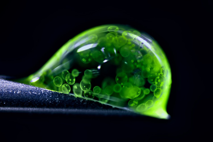
What you see here is not slime or jelly. It is a highly magnified image of a water droplet containing tiny algae balls, taken by Jan Rosenboom, a German chemical engineer, using a reflecting light microscope. Rosenboom's image won second place in Nikon's annual Photomicrograph Competition, which celebrates the contributions of microscopes to science .
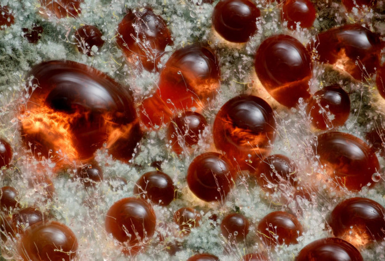
Fungi may be some of the strangest life forms on Earth, but microscopic images show just how beautiful they can be. Wim van Egmond of the Micropolitan Museum in the Netherlands took ninth place in the competition with this stunning close-up of the diffuse, red pigment of the fungus Talaromyces purpureogenus , a distant relative of the penicillin-producing fungus Penicillium .
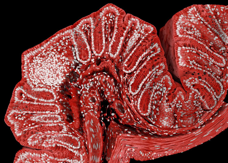
Mice play an important role in scientific research. This image of a mouse colon was taken by researchers at the Friedrich Miescher Institute for Biomedical Research in Switzerland, using magnetic resonance microscopy, a common technique in biomedical science to study cells labeled with fluorescent probes.
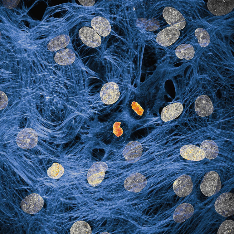
You don't have to be an expert to know that the network of cells inside our bodies works hard to make sure everything is running smoothly. James Hayes of Vanderbilt University has captured images of heart muscle cells with condensed chromosomes after cell division.
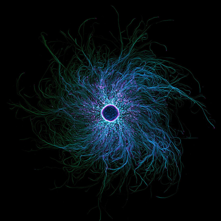
Sometimes, images of the microscopic world betray initial impressions. While this image looks a lot like a raging black hole, the subject of the photo, taken by Stella Whittaker of the National Institutes of Health (NIH), is actually sensory neurons derived from iPSCs, labeled to show two proteins, tubulin and actin. Whittaker used a combination of microscopy techniques to create this shocking image.

Filariasis is a parasitic infection caused by the specimen pictured, a parasitic roundworm. This tropical disease causes painful rashes, cell dysfunction, and even blindness. Up close, however, it doesn’t look scary at all.
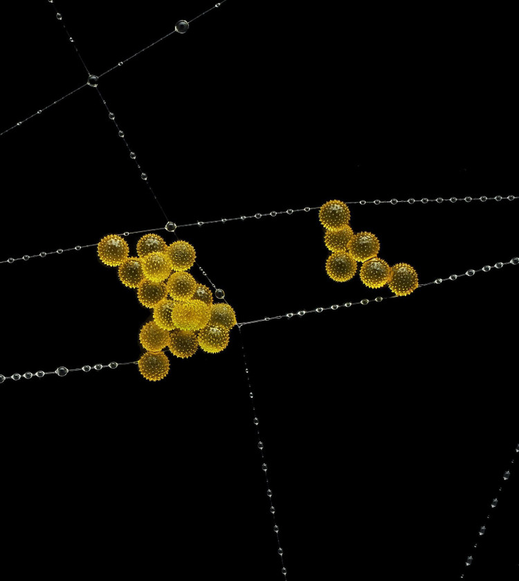
Nature is full of fragile yet incredibly resilient structures – as demonstrated by this striking image of pollen spores suspended from a garden spider web. John-Oliver Dum of Germany’s Medienbunker Produktion took third place for his image, a composite of several images stacked on top of each other.
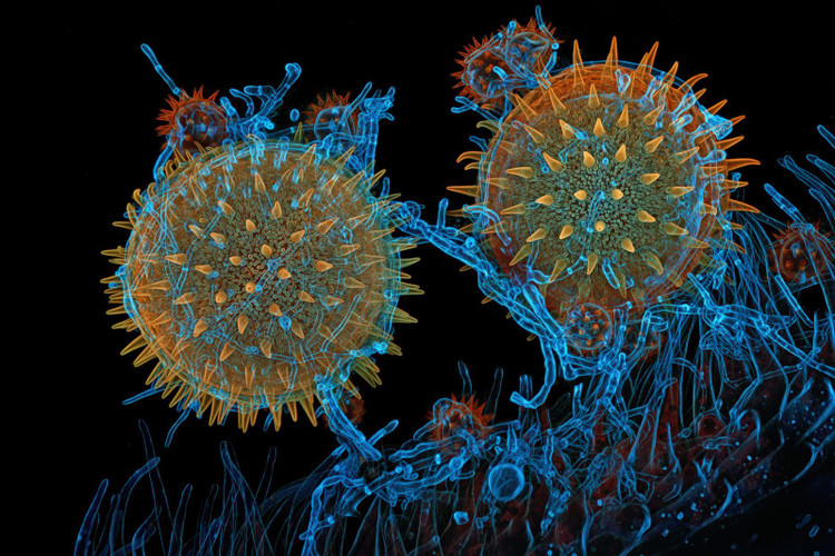
On the other hand, nature can be incredibly harsh, and extreme close-ups make that even more apparent. Igor Robert Siwanowicz of the Howard Hughes Medical Institute captured images of bone marrow pollen “germinating on a pistil while being infested with a parasitic filamentous fungus,” illustrating the wild interdependence of the microscopic world .
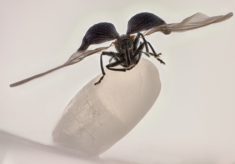
Last but certainly not least, the overall winner of the contest captured a rare moment when a rice weevil spread its wings while perched on a grain of rice. This year’s winning photo is actually a composite of over 100 images, stacked, cleaned up, and post-processed for maximum clarity and impact.
Source: https://khoahocdoisong.vn/loat-anh-hien-vi-khoa-hoc-dep-nhat-nam-don-tim-xoan-nao-nguoi-xem-post2149062463.html

![[Photo] Da Nang residents "hunt for photos" of big waves at the mouth of the Han River](https://vphoto.vietnam.vn/thumb/1200x675/vietnam/resource/IMAGE/2025/10/21/1761043632309_ndo_br_11-jpg.webp)
![[Photo] Prime Minister Pham Minh Chinh meets with Speaker of the Hungarian National Assembly Kover Laszlo](https://vphoto.vietnam.vn/thumb/1200x675/vietnam/resource/IMAGE/2025/10/20/1760970413415_dsc-8111-jpg.webp)
![[Photo] Prime Minister Pham Minh Chinh received Mr. Yamamoto Ichita, Governor of Gunma Province (Japan)](https://vphoto.vietnam.vn/thumb/1200x675/vietnam/resource/IMAGE/2025/10/21/1761032833411_dsc-8867-jpg.webp)


Comment (0)