

Standing on the brink of kidney failure because both kidneys are severely dilated
There were no typical signs, only a dull pain in the right flank, no fever, normal urination - symptoms that seemed insignificant could not fully reflect the seriousness inside the body of patient HVH (43 years old, Phu Tho ).
Upon admission, diagnostic results showed that both kidneys were in a state of severe hydronephrosis, a complication that can lead to kidney failure if not treated promptly.
Although the patient clearly felt pain in the right flank, the more serious damage was in the left kidney, where there were almost no obvious symptoms. Diagnostic imaging showed a stone in the upper third of the ureter measuring about 20x11mm and a stone in the lower renal pelvis measuring 9x6mm, causing grade IV renal pelvis dilation - the most severe level with obvious parenchymal thinning. Meanwhile, the right kidney had a stone at the junction of the renal pelvis and ureter measuring up to 25x18mm, causing grade III renal pelvis dilation.

Image of left ureteral stone causing grade IV renal pelvis dilation.
According to TCI Urology specialists, grade III and IV renal pelvis dilatation is a serious complication caused by prolonged obstruction caused by urinary stones. If not detected and treated promptly, it can lead to irreversible loss of kidney function, even chronic kidney failure.
"This is not a rare case at TCI, but the case of both kidneys being severely dilated at grade III and IV like patient H. has the potential to permanently damage both kidneys if treatment is delayed," said Meritorious Physician, Doctor CKII Pham Huy Huyen - Deputy Director of Thu Cuc International General Hospital in charge of Urology, who directly treated the case.
Not only remove the stones but also calculate to keep both kidneys
Faced with simultaneous damage to both kidneys, Meritorious Physician, Doctor CKII Pham Huy Huyen and his team chose a highly conservative treatment strategy instead of performing open surgery or simultaneous treatment.
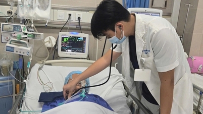
First, priority is given to treating large stones that cause obstruction at the junction of the renal pelvis and right ureter, causing pain. Percutaneous laser lithotripsy is performed to access and break up the stones, helping to clear the flow of urine.

Right renal pelvis-ureteral stones causing grade III dilatation were treated by percutaneous nephrolithotomy.
At the same time, the medical team performed left kidney drainage to release the pressure of stagnant urine, thereby minimizing further damage to kidney tissue that had been affected for a long time.
Three days after the first intervention, a second mini-tunnel lithotripsy was performed to remove the ureteral stone and the left renal calyceal stone causing grade IV renal pelvis dilatation.
"The medical team must carefully calculate so that each intervention step not only achieves the goal of cleaning the stones, but also minimizes damage to the kidney parenchyma that has been thinned due to prolonged water retention. At the same time, it does not create additional pressure on the opposite kidney during the treatment process," Dr. Huyen emphasized.
The mini-tunnel percutaneous nephrolithotomy technique is highly appreciated in the treatment of complex urinary stones thanks to its ability to precisely access and effectively remove stones with a low level of invasiveness. In particular, this technique is suitable for cases where the kidneys are damaged and need to be restored after a period of prolonged hydronephrosis.

Mini tunnel lithotripsy technique is as small as a chopstick tip, preserving the kidney.
From alarmingly severe dilation to marked improvement after one month
Thanks to the application of modern techniques, both interventions were performed safely, the patient recovered well and was discharged from the hospital 3 days after the second lithotripsy.
After about a month, the examination results showed significant improvements. The bilateral renal pelvis dilation had reduced to grade II, instead of grades III and IV as before. The kidney size returned to normal, the renal parenchyma was clearly differentiated between the medulla and cortex. This was a positive sign that the urine flow had been released, the kidney began to gradually recover after the cause of obstruction was removed, and renal function was still well preserved.

Patient H. had no stones, severe dilated renal pelvis improved.
The case of HVH patient also shows a remarkable fact, urinary stones can progress silently, without showing obvious symptoms but causing serious damage to the kidneys. If detected late, complications may no longer be able to recover. Therefore, regular check-ups and timely treatment intervention when there are abnormal signs play an important role in protecting the health of the kidneys and urinary system.
Thu Cuc International General Hospital masters advanced urinary kidney stone lithotripsy technologies, minimizing invasion and preserving kidney function. TCI currently offers up to 30% discount on lithotripsy costs for all methods.
For more information, visit https://benhvienthucuc.vn/tan-soi-cong-nghe-cao-danh-bay-soi-tiet-nieu/
Contact: 1900 55 88 92.
Source: https://dantri.com.vn/suc-khoe/cuu-hai-qua-than-khoi-bo-vuc-suy-than-tu-bien-chung-gian-nang-20250515184829864.htm

![[Photo] General Secretary To Lam attends the National Conference to disseminate and implement 4 Resolutions of the Politburo](https://vphoto.vietnam.vn/thumb/1200x675/vietnam/resource/IMAGE/2025/9/16/70c6a8ceb60a4f72a0cacf436c1a6b54)
![[Photo] General Secretary To Lam chaired a working session with the Standing Committee of the Party Committee of the Ministry of Foreign Affairs](https://vphoto.vietnam.vn/thumb/1200x675/vietnam/resource/IMAGE/2025/9/15/f26e945b18984e8a99ef82e5ac7b5e7d)
![[Photo] National conference to disseminate and implement 4 Resolutions of the Politburo](https://vphoto.vietnam.vn/thumb/1200x675/vietnam/resource/IMAGE/2025/9/16/5996b8d8466e41558c7abaa7a749f0e6)

![[Photo] Prime Minister Pham Minh Chinh attends the closing ceremony of the exhibition of national achievements "80 years of the journey of Independence - Freedom - Happiness"](https://vphoto.vietnam.vn/thumb/1200x675/vietnam/resource/IMAGE/2025/9/15/a1615e5ee94c49189837fdf1843cfd11)


Comment (0)