
NDO – On December 2, the Maxillofacial Department (B8), Military Hospital 175 successfully performed the first surgery to resect the lower jaw bone and reconstruct it with a free peroneal muscle flap, applying modern 3D technology. This is a breakthrough in the treatment of ameloblastoma, bringing about results beyond expectations in both function and aesthetics for the patient.
Male patient NQN (1995, Quang Ngai ) discovered a tumor in the lower jaw bone 6 years ago but stopped treatment after the initial follow-up period. Recently, the swelling increased, the patient came to Military Hospital 175 with a swollen lower jaw, mild red inflammation of the mucosa, and little pain. The results of the diagnostic imaging showed that the tumor destroyed the bone from tooth number 44 to the ascending branch of the right lower jaw.
Ameloblastoma is a benign tumor but has the ability to destroy bone and has the potential to become malignant if not treated promptly. Surgery to remove the jawbone to remove the tumor often causes serious functional and aesthetic losses, greatly affecting the patient's quality of life, especially in young people.
The patient underwent surgery to resect the mandible, remove the tumor with a 1cm safe margin; at the same time, reconstruct the mandible with a free peroneal musculocutaneous flap.
This is a complex technique, requiring skill, meticulousness and endurance from the surgical team. However, this method is supported by 3D technology, helping doctors accurately simulate each cutting, grafting and reconstruction operation. Thanks to that, the surgical results are optimized in both function and aesthetics, bringing outstanding efficiency to the patient.
The patient is designed with a cutting line and surgical guide to cut the jawbone and fibula (cutting guide) on the software, the guide is printed, and a 3D jaw model is printed after simulating bone cutting and reconstruction.
Bend the splint according to the simulated jaw model first. Then, cut the mandibular bone segment from the high branch to the distal side of tooth 42. The dissected fibula muscle flap will be cut according to the cutting guide.
The fibula is incorporated into the pre-bent reconstruction splint, then the flap complex is incorporated, and the splint is placed into the mandible to ensure occlusion and jaw mobility. Finally, microsurgical vascular anastomosis ensures the survival of the reconstruction flap.
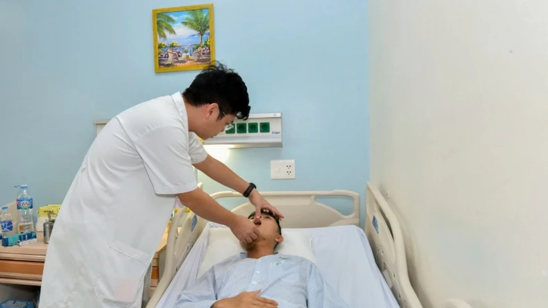 |
After 10 days of surgery, the patient recovered well: stable overall, no fever, normal vital signs, good fibula flap, dry surgical wound, fast healing, eating, chewing, opening and closing function and facial aesthetics were significantly improved. |
The lower jaw not only plays an important role in chewing, speaking, and swallowing, but also shapes the facial structure.
Therefore, the reconstruction of large defects after surgery is always a top priority. The free fibula flap with 3D printing technology brings many advantages: Ensuring enough bone supply for the reconstruction of large segments, allowing two surgical teams to perform simultaneously to shorten the surgical time and increase the accuracy and effectiveness of treatment.
Doctor Do Van Tu, Department of Maxillofacial Surgery, Military Hospital 175, who directly performed the surgery, said: "Using software to create a "cutting guide" and print a jaw model helps surgeons have the most precise cutting lines, especially helping to create the most perfect lower jaw bone from the fibula from every angle.
Calculating the details of the cuts and grafts will help doctors perform surgery simply and accurately, instead of having to wait until the patient is on the operating table to perform time-consuming calculations like before.
Proper planning and execution of the plan when applying 3D technology in microsurgery is an important factor to ensure optimal aesthetic and functional results for the patient.”
Nowadays, 3D technology is applied more and more popularly in the field of Dentistry such as orthopedic surgery, dental implant surgery, plastic surgery...
Therefore, using 3D technology to design simulations and plan microsurgical mandibular flap surgery helps to simplify, not only improve accuracy and shorten surgery time, but also optimize treatment results, especially in complex cases such as mandibular reconstruction.
The combination of 3D printing technology and microsurgery techniques is a testament to the continuous efforts of Military Hospital 175 in applying advanced technology, providing optimal solutions, meeting treatment needs and improving the quality of life for patients.
![[Photo] Editor-in-Chief of Nhan Dan Newspaper Le Quoc Minh received the working delegation of Pasaxon Newspaper](https://vphoto.vietnam.vn/thumb/1200x675/vietnam/resource/IMAGE/2025/9/23/da79369d8d2849318c3fe8e792f4ce16)

![[Photo] Prime Minister Pham Minh Chinh chairs the 14th meeting of the Steering Committee on IUU](https://vphoto.vietnam.vn/thumb/1200x675/vietnam/resource/IMAGE/2025/9/23/a5244e94b6dd49b3b52bbb92201c6986)
![[Photo] The 1st Congress of Party Delegates of Central Party Agencies, term 2025-2030, held a preparatory session.](https://vphoto.vietnam.vn/thumb/1200x675/vietnam/resource/IMAGE/2025/9/23/e3a8d2fea79943178d836016d81b4981)

![[Photo] General Secretary To Lam meets voters in Hanoi city](https://vphoto.vietnam.vn/thumb/1200x675/vietnam/resource/IMAGE/2025/9/23/d3d496df306d42528b1efa01c19b9c1f)


Comment (0)