
When the pregnancy was 25 weeks and 4 days, pregnant woman NT T (30 years old, from Thanh Hoa) came to Hanoi Obstetrics Hospital for a check-up in a worried state because before that, at the 22nd week of pregnancy, she was discovered to have a pleural effusion in the right pleura during a check-up at a local hospital.
But after only a few weeks, the condition progressed rapidly - pleural effusion increased significantly on both sides, Ms. T. was referred to Hanoi Obstetrics Hospital for in-depth examination.
At the Fetal Intervention Center, Hanoi Obstetrics Hospital, ultrasound results showed that the fetus had bilateral pleural effusion, the right side was 70mm thick, the left side was 38mm thick; almost all of the right lung parenchyma and part of the left lung parenchyma collapsed. The fetus had polyhydramnios and the heart was displaced due to compression by the fluid.
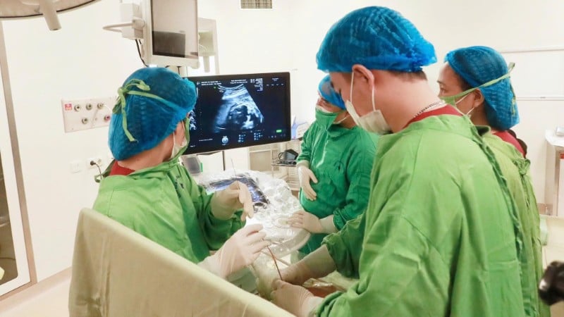
Doctors determined that this was a case of severe pleural effusion, causing lung collapse and risk of pulmonary hypoplasia, requiring early intervention to reduce pressure in the fetal chest. After consultation, the Center team decided to perform fetal intervention to place a shunt to drain the pleural fluid.

Under ultrasound guidance, a guide needle is gently inserted through the mother’s abdominal wall, reaching the fetal pleural cavity. The tiny shunt is placed in the appropriate position, helping to drain fluid from the pleural cavity out of the amniotic sac, reducing pressure and allowing the fetal lungs to re-expand.
At the same time, excess amniotic fluid is also removed to ensure it is appropriate for the current gestational week.
Three days after the intervention, the ultrasound results showed that the pleural fluid was gradually draining out, the left lung no longer had fluid and the right lung began to expand well, the fetal heart was no longer displaced as before, the abdominal fluid had completely disappeared and there was no longer excess amniotic fluid.
When seeing the image of the tiny shunt on the ultrasound screen, the mother felt like she saw another ray of hope - proof of the miraculous recovery of the fetus that is taking place every day in the mother's womb.
Source: https://nhandan.vn/hoi-sinh-cho-thai-nhi-25-tuan-tuoi-bi-tran-dich-mang-phoi-hai-ben-post924176.html


![[Photo] The Standing Committee of the Organizing Subcommittee serving the 14th National Party Congress meets on information and propaganda work for the Congress.](https://vphoto.vietnam.vn/thumb/1200x675/vietnam/resource/IMAGE/2025/11/19/1763531906775_tieu-ban-phuc-vu-dh-19-11-9302-614-jpg.webp)


![[Photo] Prime Minister Pham Minh Chinh and his wife meet the Vietnamese community in Algeria](https://vphoto.vietnam.vn/thumb/1200x675/vietnam/resource/IMAGE/2025/11/19/1763510299099_1763510015166-jpg.webp)
![[Photo] General Secretary To Lam receives Slovakian Deputy Prime Minister and Minister of Defense Robert Kalinak](https://vphoto.vietnam.vn/thumb/1200x675/vietnam/resource/IMAGE/2025/11/18/1763467091441_a1-bnd-8261-6981-jpg.webp)


Comment (0)