
Determined to keep the pregnancy for the pregnant woman
The 37-year-old patient, NTH, had undergone surgery to treat endometriosis and uterine fibroids. After surgery, natural pregnancy became more difficult for Ms. H. due to severe ovarian reserve reduction. Ms. H. had to undergo assisted reproduction with a small number of embryos to get pregnant, but unfortunately, the fetus had Trisomy 18 (Edwards Syndrome) and had to be terminated at week 19.
After many efforts, she became pregnant with baby G. At the 17th week, when performing a morphological ultrasound at the Fetal Medicine Center, Tam Anh General Hospital, Ho Chi Minh City, it was discovered that the fetus had a tumor in the sacral region. At this time, the tumor was about 3-4cm long.
After consulting with doctors, the couple decided to keep the pregnancy, even though about 17% of sacral teratoma have malignant factors that cannot be examined in the fetus.
At 22 weeks of pregnancy, the fetal MRI results showed a large tumor, rapidly increasing in size, many blood vessels proliferating in the tumor, and the tumor growing outside the body. By the 30th week, the tumor size increased 5 times, with a risk of fetal anemia, heart failure, tumor rupture, fetal death, etc.
The doctor determined that the fetus had a type 1 sacral teratoma. The tumor grew completely outside the body with many proliferating blood vessels. A large amount of blood from the fetal circulation went into the tumor, causing the fetus to gradually become anemic and lead to heart failure.
By the 34th week of pregnancy, the tumor has grown to twice the size of the baby's body, and the fetus is showing signs of heart failure. If the tumor ruptures, it will lead to fetal hemorrhagic shock, threatening the lives of both mother and baby.
The Obstetrics-Neonatal and Pediatric Surgery departments plan to coordinate pregnancy monitoring, cesarean section, postnatal care, and surgery when the baby is healthy enough.
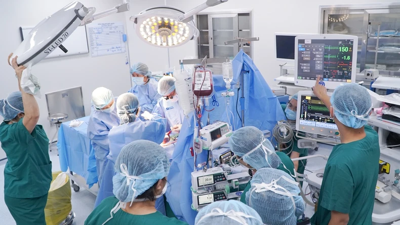
Two adjacent operating rooms were prepared for the worst case scenario, one with a team of obstetricians and neonatologists to perform a cesarean section and provide initial care for the mother and child, the other with a team of pediatricians and anesthesiologists to perform the tumor removal surgery.
Two adjacent operating rooms are prepared for the worst case scenario, |
Four hours of brain surgery to remove the tumor, saving the life of the fetus
Specialist II Doctor Nguyen Ba My Nhi, Director of the Obstetrics and Gynecology Center of Tam Anh General Hospital in Ho Chi Minh City, directly performed the cesarean section and said that if this case continued to drag on, the fetus could die suddenly. If the tumor ruptured in the uterus, it would cause a lot of blood loss and threaten the lives of both mother and child.
During the cesarean section, the surgical team faces the risk of rupturing the tumor when removing the fetus from the uterus, causing severe bleeding that may not be able to be stopped, leading to the baby's immediate death due to acute massive blood loss.
Therefore, the doctors decided to make a large vertical incision from the lower segment to the bottom of the uterus, carefully removing the fetus safely without rupturing the tumor. The mother's uterus was quickly restored by sutures, no postpartum hemorrhage occurred, and no blood transfusion was needed.
Baby G. was born safely at the end of April 2025, weighing 3.4 kg, of which the tumor weighed 1.8 kg and was nearly 20 cm long. After birth, the neonatologists gave the baby oxygen, provided postnatal care, and stabilized her vital signs. The pediatrician examined her and decided to take her to the Neonatal Intensive Care Unit (NICU). It is expected that after 24 hours, when the baby is stable, surgery will be performed to remove the tumor.
Two hours later, bleeding began inside the tumor, the tumor grew larger, and the child was at risk of hemorrhagic shock, threatening his life. The hospital immediately activated the emergency mode for the entire hospital and transferred the child to the operating room.
The pediatric surgeon team including specialist II Nguyen Do Trong, Master, doctor Lam Thien Kim and Master, specialist I Ton Thi Anh Tu immediately appeared with neonatal anesthesiologists to perform surgery to stop the bleeding and remove the tumor for the baby.
Doctor Trong said that the tumor was attached to the body, if separated it would cause the child to lose a large amount of blood, so both resuscitation and surgery were required. The team of anesthesiologists ensured hemodynamics, and the surgeon controlled bleeding. At the same time, the patient was given a blood bank, and all blood products such as red blood cells, plasma, platelets, etc. were prepared to compensate for the child during and after surgery.
Assessing that the tumor is very large, possibly related to the colon, bladder, genitourinary organs and surrounding structures, doctors need to completely separate the tumor, limit blood loss, damage to surrounding organs (perforation of the rectum, vagina, etc.), maximally protect the anal sphincter and nerves of the perineum, ensuring the child's aesthetics.
After nearly 4 hours, the tumor was removed from the body and the patient was transferred to the NICU for continued monitoring.
24 hours after the cesarean section, the health of both mother and child was stable. The mother could eat, drink, move around, the uterus contracted well and there was no vaginal bleeding. The baby's circulation and breathing were stable, and he was taken off the ventilator after a day, and the incision was no longer bleeding. During and after the surgery, the baby received a 500ml blood transfusion.
Dr. Cam Ngoc Phuong, Director of the Neonatal Center, said the baby continued to be monitored, and in mid-May, the baby's health stabilized and he was discharged from the hospital.
Sacrococcygeal teratoma is a rare disease with a rate of about 1/20,000-40,000 births, divided into 4 types. The cause of the disease is currently unknown. If not treated promptly, the disease can cause fetal heart failure in the baby, and in the mother it can cause polyhydramnios and mirror rotation syndrome, leading to placental edema. In particular, the tumor can rupture at any time before, during and after birth, causing both mother and baby to go into hemorrhagic shock, leading to death.
Sacrococcygeal teratoma can be detected early through prenatal ultrasound using modern diagnostic imaging methods, thereby assessing the risk and developing treatment plans for the child as soon as it is born.
Source: https://nhandan.vn/cuu-be-so-sinh-mang-u-quai-gan-2kg-post880768.html

![[Photo] Top players gather at the 2025 Nhan Dan Newspaper National Table Tennis Championship](https://vphoto.vietnam.vn/thumb/1200x675/vietnam/resource/IMAGE/2025/5/23/9ad5f6f4faf146b08335e5c446edb107)


![[Video] Ho Chi Minh City records an increase in Covid-19 cases](https://vphoto.vietnam.vn/thumb/402x226/vietnam/resource/IMAGE/2025/5/23/fcd232389af14440b901b7e2cd0982dc)


![[Photo] Top players gather at the 2025 Nhan Dan Newspaper National Table Tennis Championship](https://vphoto.vietnam.vn/thumb/402x226/vietnam/resource/IMAGE/2025/5/23/9ad5f6f4faf146b08335e5c446edb107)


Comment (0)