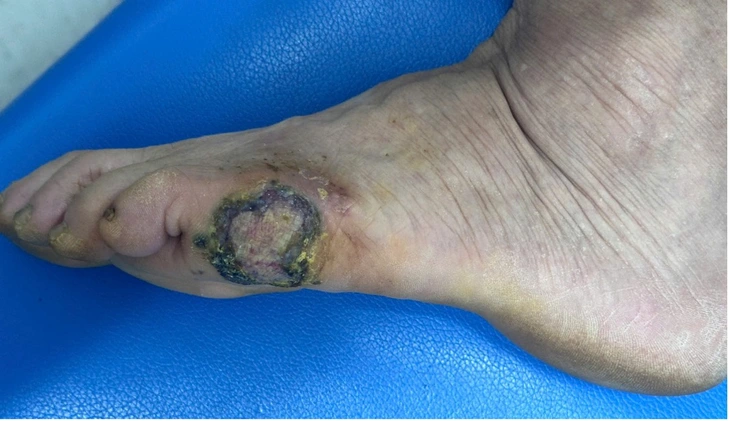
Image of black patch on woman's foot - Photo: BVCC
According to the patient, this black patch appeared many years ago. About a year ago, she saw the black patch gradually getting bigger but it was not painful or itchy.
She then went to a private clinic and had the entire lesion removed. About 3 months after the procedure, the old lesion began to reappear with a black spot that grew larger and had a bleeding ulcer in the center.
After examination, Dr. Vu Nguyen Binh, Department of Plastic Surgery and Rehabilitation, Central Dermatology Hospital, assessed that the patient was alert, had no fever, and was in stable condition.
The main lesion is a hyperpigmented patch on the outer edge of the left foot, measuring 4x3cm, with a relatively clear boundary compared to the surrounding healthy skin, uneven edges, and a dirty scaly surface. The patient also has some diseases such as high blood pressure, kidney stones, etc.
Dr. Binh ordered the patient to undergo a dermoscopy of the lesion, a specialized paraclinical test in dermatology. The results of the scan matched the doctor's clinical diagnosis of melanoma and the patient was admitted to the hospital for treatment.
According to Dr. Binh, the lesion appeared and progressed over a long period of time. The patient and his family subjectively did not go to the doctor early because they thought it was a normal, non-dangerous mole.
The patient only goes to the doctor when the lesion is too large and starts bleeding. At this point, the lesion may have progressed to cancer.
In addition, patients are first treated at non-specialized facilities, so the risks cannot be fully assessed and the treatment method is incorrect.
At the Central Dermatology Hospital, the patient was prescribed a wide excision of the lesion (2cm from the edge of the lesion). Because the lesion was located close to the little toe, to ensure safety, the doctor performed surgery to amputate the little toe of the left foot.
The lesion is then examined by pathology to determine the depth of invasion of cancer cells to assess metastasis.
In addition, patients are also screened for metastasis by sentinel lymph node biopsy, which is a valuable initial metastasis screening method.
Fortunately for the patient, the cancerous lesion had not invaded too deeply in the affected area and there were no signs of metastasis to the lymph nodes. The patient's prognosis was relatively good, and the patient was then given a skin graft to cover the skin defect after the cancerous lesion was removed.
This method helps restore lost skin, and makes it easier to monitor for recurrence after treatment.
Look out for brown, black lesions.
Dr. Nguyen Hong Son, head of the Department of Plastic Surgery, Central Dermatology Hospital, advised that every year the hospital receives many patients with injuries and diagnoses similar to this patient.
"Therefore, when detecting hyperpigmented lesions (brown, black) on the palms of the hands and soles of the feet, people need to immediately go to specialized facilities for early detection and timely treatment, minimizing possible unfortunate consequences, because hyperpigmented lesions in these areas have a high cancer rate and high malignancy level," Dr. Son warned.
Source: https://tuoitre.vn/phat-hien-mac-ung-thu-tu-mang-den-o-ban-chan-2025051217510585.htm


![[Infographic] In 2025, 47 products will achieve national OCOP](https://vphoto.vietnam.vn/thumb/402x226/vietnam/resource/IMAGE/2025/7/16/5d672398b0744db3ab920e05db8e5b7d)


Comment (0)