
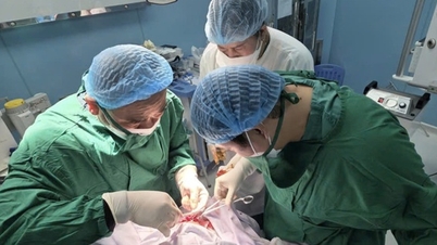
The tumor was carefully dissected and successfully removed from the patient's brain after 4 hours without causing damage to the surrounding brain tissue. The patient made a spectacular recovery just 2 hours after surgery.
Stroke screening, unexpectedly discovered large tumor compressing brain tissue
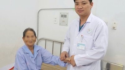
Ms. Trinh Nhat Khanh (a 45-year-old female martial artist living in Ho Chi Minh City) occasionally suffered from mild headaches, so she decided to go for a stroke screening at FV Hospital. The MRI results unexpectedly discovered a very large meningioma , more than 6 cm in diameter, looking like an orange, compressing nearly 1/4 of the brain.
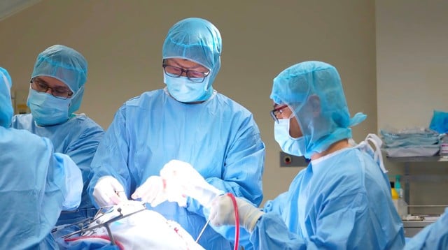
The tumor was successfully removed after 4 hours without causing damage to the surrounding brain tissue. The patient recovered only 2 hours after surgery.
Photo: BVCC
"The tumor develops over a long period of time, the skull adapts to the tumor so it is difficult to detect. The tumor has not affected higher neurological functions, although the patient occasionally has headaches. However, this case needs to be operated on early, because if the tumor gets larger, it can cause dangerous complications such as epilepsy or even directly threaten the patient's life, " said specialist doctor Tran Luong Anh, Head of the Department of Neurosurgery and Spine.
From the consultation between the Department of Diagnostic Imaging and the Department of Neurosurgery and Spine, the doctors determined that for the surgery to be successful, two major challenges need to be solved.
First, the tumor is large and hard, so removing it can cause damage to the surrounding brain tissue. Second, the tumor is nourished by many blood vessels, so there is a risk of heavy blood loss during surgery.
Interventional and surgical intervention with Navigation system, complete tumor resection, brain tissue preservation
To minimize blood loss during surgery and have a plan to preserve blood vessels , Dr. Luong Anh asked specialist Dr. Huynh Huu Danh, a vascular intervention specialist, to perform an embolization procedure, blocking the blood supply to the tumor one day before surgery . Embolization, according to Dr. Danh, is a minimally invasive method used to treat a number of brain diseases such as cerebral vascular malformations, cerebral aneurysms, carotid-cavernous sinus fistulas, dural arteriovenous fistulas, etc.
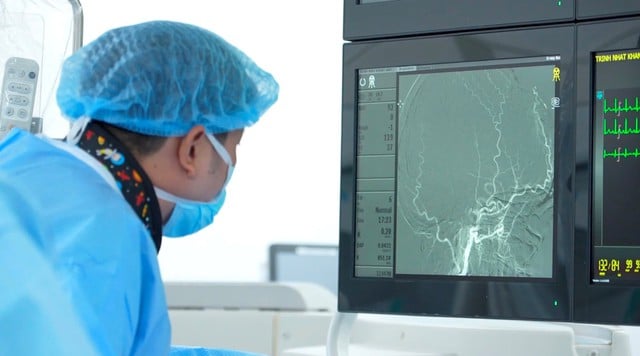
Image of cerebral blood vessels around the tumor during embolization
Photo: BVCC
Doctor Huynh Huu Danh performed vascular intervention, the procedure took about 30 minutes, 90% of the blood vessels feeding the tumor were completely blocked.
Day The next day, the surgery, led by Dr. Luong Anh, took place under the guidance of neuronavigation . This system helps to accurately determine the tumor's boundaries, optimize the incision so that it is not too large or too small, thereby reducing the surgery time and related risks. Navigation also helps to identify and preserve the blood vessels adjacent to the tumor.
" With large and hard tumors like this, removing the tumor manually can cause damage to the surrounding brain tissue. The ultrasonic cutter helps remove the tumor without moving it much, minimizing the impact on brain tissue and protecting the surrounding blood vessels, " Dr. Luong Anh explained .
The tumor was carefully dissected and successfully removed from the patient's brain after 4 hours without causing damage to the surrounding brain tissue. Thanks to the coordination with the neurovascular interventionalist to block the tumor's blood supply , the amount of bleeding during surgery was very little, the surgery time was shortened, and the patient recovered spectacularly just 2 hours after surgery.
Source: https://thanhnien.vn/boc-tron-khoi-u-nao-6-cm-benh-nhan-hoi-phuc-sau-2-gio-phau-thuat-185250730161517417.htm
![[Photo] Panorama of the 2025 Community Action Awards Final Round](https://vphoto.vietnam.vn/thumb/1200x675/vietnam/resource/IMAGE/2025/11/15/1763206932975_chi-7868-jpg.webp)

![[Photo] Prime Minister Pham Minh Chinh meets with representatives of outstanding teachers](https://vphoto.vietnam.vn/thumb/1200x675/vietnam/resource/IMAGE/2025/11/15/1763215934276_dsc-0578-jpg.webp)

![[Photo] General Secretary To Lam receives Vice President of Luxshare-ICT Group (China)](https://vphoto.vietnam.vn/thumb/1200x675/vietnam/resource/IMAGE/2025/11/15/1763211137119_a1-bnd-7809-8939-jpg.webp)


Comment (0)