
To detect and treat mediastinal tumors early, people should have regular health check-ups and perform tests such as chest X-rays or chest CT scans as prescribed by their doctors.
Medical news on January 15: Warning about mediastinal cancer through two cases and advice from doctors
To detect and treat mediastinal tumors early, people should have regular health check-ups and perform tests such as chest X-rays or chest CT scans as prescribed by their doctors.
Warning signs of mediastinal cancer
One day at the end of 2024, Mr. M. (42 years old, Ho Chi Minh City) went to Cho Ray Hospital for examination because of prolonged hoarseness. The results of a full-body CT scan surprised him when it discovered a thymic tumor in the middle mediastinum, measuring 60x36mm.
After surgery and pathology, the doctor confirmed that he had thymic carcinoma. Immediately, the doctors consulted and gave him emergency treatment solutions.
Meanwhile, Ms. T. (32 years old, Hanoi ) also went to Cho Ray Hospital for examination when she had a bad cough and chest pain. The chest CT scan results showed that she had a superior anterior mediastinal tumor measuring 68x43mm, along with many enlarged lymph nodes in the mediastinum and left lung hilum. After a biopsy, the results showed that she had lymphoma. However, because this type of cancer often responds well to chemotherapy, her condition has gradually stabilized, and she can return to work normally.
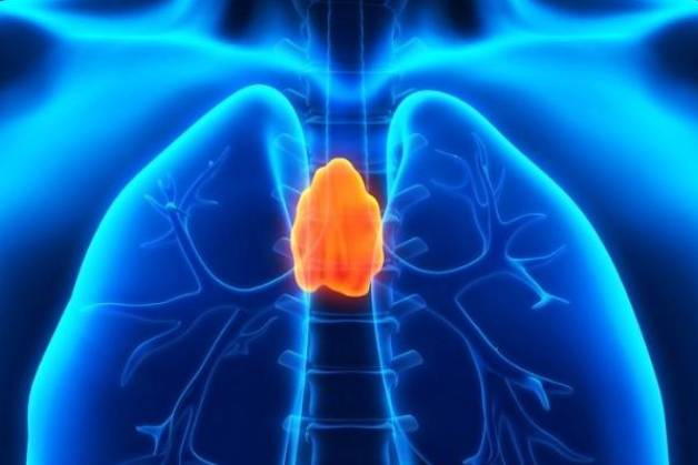 |
| The mediastinum is the central region of the chest, which contains many important organs such as the heart, esophagus, trachea, large blood vessels, nerves, and thymus gland. Illustration photo |
According to specialist 2 Cao Thi Hong, Head of HECI Center, Cho Ray Hospital, the mediastinum is the central region of the chest, which contains many important organs such as the heart, esophagus, trachea, large blood vessels, nerves and thymus. Tumors here can be benign or malignant, including thymoma, thymoma, lymphoma, germ cell tumor, neuroma and many other types.
Although mediastinal tumors often develop silently in the early stages, when patients experience symptoms such as chest pain, difficulty breathing when lying on their back, hoarseness, severe cough or difficulty swallowing, the disease has entered a late stage and can be life-threatening. Treatment of mediastinal tumors is complex and requires a combination of surgery, chemotherapy, and radiotherapy and should be performed at specialized general hospitals.
Doctors recommend that, in order to detect and treat mediastinal tumors early and promptly, people should have regular health check-ups and perform tests such as chest X-rays or chest CT scans as prescribed by their doctors. Early detection and intervention can significantly reduce the risk of death and give patients a better chance of effective treatment.
Revolution in the treatment of neurological diseases
The remarkable development of science and technology, especially artificial intelligence, is opening up great opportunities in the field of medicine, especially in the treatment of neurological diseases. This information was shared by Lieutenant Colonel, Dr. Nguyen Xuan Phuong, Deputy Head of the Department of Neurosurgery, Military Hospital 103, at the "2024 Military-Political Conference" recently held at this hospital.
Dr. Phuong pointed out the challenges in treating brain tumors, especially for tumors deep in the brain, where there is no clear boundary with the brain parenchyma. Complete tumor removal surgery, while minimizing complications and preserving brain function, is always a difficult problem. To solve this problem, surgical planning using artificial intelligence, using surgical robotic systems combined with CT and MRI technology during surgery is being developed and applied in hospitals.
According to Dr. Phuong, the Department of Neurosurgery at Military Hospital 103 has implemented many new techniques, bringing high efficiency in treating patients. One of the typical techniques is open craniotomy combined with opening the cerebrospinal fluid reservoir at the base of the skull to treat severe traumatic brain injury. This is the first technique performed in Vietnam, aiming to adjust intracranial pressure in patients with severe traumatic brain injury.
In addition, endoscopic surgery to remove hematomas in the brain after stroke is also widely applied, helping to improve the effectiveness of treatment for stroke patients. In particular, in these surgeries, intelligent software is used to ensure thorough removal of hematomas and preservation of brain function.
Lieutenant General, Associate Professor, Dr. Nghiem Duc Thuan, Political Commissar of the Military Medical Academy, commented that 2024 will be a challenging year for Military Hospital 103. The hospital will simultaneously carry out many important tasks, from medical examination and treatment, scientific research to ensuring national security and defense. Medical examination and treatment at the hospital has applied many modern technologies, especially in the field of organ transplantation.
"In particular, many highly applicable research topics have been implemented, helping to solve practical problems, especially in the strong development of scientific research on nursing. Thanks to that, the hospital has received trust and affection from patients and their families," Associate Professor, Dr. Thuan shared.
The Political Commissar of the Military Medical Academy also requested that Military Hospital 103 continue to improve medical examination and treatment processes, promote digital transformation, apply science and technology, and develop new techniques in 2025.
Treatment of infected coral kidney stones with modern endoscopic surgery
Ms. NTTV, 53 years old, from Khanh Hoa, has been experiencing pain and numbness in her lower back for the past two months without a clear cause. The pain often occurs when she bends over or works, accompanied by a feeling of fatigue, easy hip shock, and having to lie on her right side to relieve the pain. Recently, she also discovered that her urine was cloudy and had an unpleasant odor, so she decided to go to Tam Anh General Hospital in Ho Chi Minh City for a check-up.
At the hospital, Dr. Nguyen Truong Hoan, Department of Urology, Center for Urology - Nephrology - Andrology, ordered her to undergo a computed tomography (CT) scan to examine the lumbar region. The results showed that her left kidney showed signs of hydronephrosis, inside the renal pelvis there was a large stone with 4 branches, penetrating into the renal calyces. The upper and middle calyces of the kidney also contained many smaller stones, with a total size of up to 5-6cm, accounting for about ⅓ of the volume of the left kidney. In addition, she also had a urinary tract infection.
According to Dr. Hoan, Ms. V. suffered from infected coral stones, a dangerous type of kidney stone that required surgery to remove the stones and restore urinary flow. Before surgery, the patient was treated with antibiotics for 1 week, then had a urine culture done to ensure the infection was under control. Treatment of urinary tract infections is very important, because if left unchecked, bacteria from the stones can enter the bloodstream, threatening the patient's life during endoscopic lithotripsy.
After the urinary tract infection stabilized and the urine culture results were negative, Ms. V. was treated with percutaneous endoscopic lithotripsy (mini PCNL), which is currently the optimal technique for large coral stones. Compared with open surgery, this method has outstanding advantages such as less bleeding, less risk of surgical site infection and less pain, helping patients recover quickly.
During the surgery, the medical team uses ultrasound and the C-Arm system to precisely locate the stone and insert a needle through a small tunnel less than 1cm from the skin into the renal pelvis. The stone is then broken into small pieces by high-power laser energy and suctioned out.
After only 180 minutes of lithotripsy, the entire coral stone mass was removed from Mrs. V's left kidney. After 1 day of recovery, she was discharged from the hospital because she was no longer in pain and could eat and walk normally. A week later, the ultrasound results of her left kidney showed that there were no more stones and her health was stable.
Coral stones account for only about 10%-15% of urinary stones but are the most dangerous type. These stones often develop rapidly in an environment with urinary tract infections, which can lead to hydronephrosis and cause serious damage to kidney function. Women are twice as likely to develop coral stones as men due to their susceptibility to urinary tract infections.
Doctor Hoan said that coral stones develop silently, often with few or mild symptoms such as lower back pain, cloudy urine, fatigue, etc. If not detected and treated promptly, coral stones can cause kidney infections, pyelonephritis, kidney failure or life-threatening blood infections.
Therefore, doctors recommend that people with similar symptoms, especially those with a history of coral stones, should go to the hospital early for examination and timely treatment. In addition, it is advisable to develop a habit of regular health check-ups every 6-12 months to detect stones when they are still small, allowing for treatment with minimally invasive methods such as extracorporeal shock wave lithotripsy or medication.
Source: https://baodautu.vn/tin-moi-y-te-ngay-151-canh-bao-ung-thu-trung-that-qua-hai-ca-benh-va-loi-khuyen-tu-bac-sy-d240799.html


Comment (0)