
That is Mr. PVĐ (69 years old, residing in Vi Thanh commune, Can Tho city), with a history of poorly controlled diabetes.
About two weeks before being admitted to the hospital, Mr. D. had a prolonged migraine, accompanied by reduced vision in his right eye. After that, his vision rapidly decreased and was completely lost while his left eye still had normal vision.
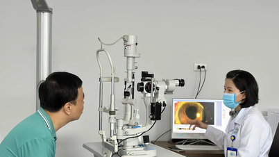
Mr. D. visited several medical facilities with ophthalmology departments, but no abnormal signs of eye damage were found. After further examinations at several other medical facilities, he was diagnosed with a brain tumor. Therefore, his family transferred him to SIS Can Tho International General Hospital for examination and monitoring.
Hidden sinus mass is easily confused
Here, doctors noted that the patient's right eye no longer responded to light, but eye structures such as the cornea, conjunctiva, retina and eyeball movements were still normal.
This shows that the damage is most likely located on the optic nerve pathway, not an eye disease. Therefore, Mr. D. was ordered to have an MRI scan of the brain and cerebral vessels to rule out causes in the brain parenchyma.

The deep sinus mass is destroying the skull base.
MRI results showed no hemorrhage, infarction, or brain tumor. However, an abnormal mass appeared in the optic nerve canal area right next to the lateral wall of the sphenoid sinus, compressing the optic nerve. Based on this suspicion, the doctors ordered a CT scan to determine the nature of the injury.
According to Dr. Vo Van Nam, resident physician specializing in ENT, CT-Scan images show many typical signs of sphenoid sinus fungus.
"The sinus wall has a thick, destroyed area, with increased luminosity in the sinus cavity and density consistent with fungal lesions. The fungal mass has invaded the optic nerve, which is the direct cause of vision loss" - Dr. Nam analyzed.
According to experts, the sphenoid sinus area is located right next to the base of the skull, so it is easy to be confused when taking an MRI. Therefore, clinical experience is essential to recognize this situation. "Sphenoid sinus fungal lesions can be mistaken for brain tumors," Dr. Nam noted.
Overcoming difficulties to restore vision
After consultation, the team of doctors assessed that the fungal mass was located close to the internal carotid artery. This is an important blood vessel branch that nourishes the brain. At the same time, the fungal mass had eroded the skull base bone, making the surgery difficult and potentially risky.

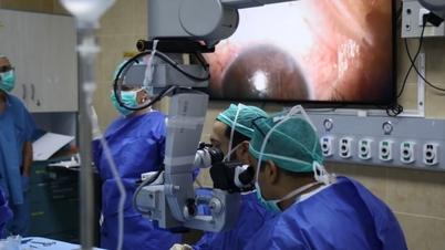
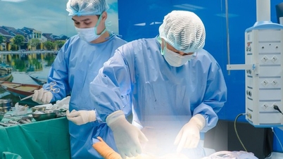
The surgical team is struggling to handle Mr. D's situation.
To ensure maximum safety, the surgical team uses a 3D Navigation system combined with magnifying endoscopy to accurately determine the location of the fungal nest and the boundary of healthy tissue.
"We opened the sphenoid sinus, decompressed the optic nerve and removed the entire mass of fungus. Fortunately, the meninges were still intact, which significantly reduced the risk of complications such as cerebrospinal fluid leakage or carotid artery damage," Dr. Nam added.
According to experts, surgeries in special locations like this case, without a 3D positioning system and precise techniques, patients may face dangerous bleeding, cerebrospinal fluid leakage, or damage to important nerve structures.
Just 24 hours after surgery, the patient began to perceive light and distinguish human shadows. "If it had been later, the optic nerve could have been irreversibly damaged, leading to permanent blindness," the expert added.
Test results showed that the causative agent was Aspergillus fumigatus, a fungus commonly found in people with diabetes or immunodeficiency.
Notes for people with diabetes
Experts warn that people with poorly controlled diabetes are susceptible to fungal infections, including sphenoid sinus fungus. Sphenoid sinus fungus often presents discreetly and is easily confused with other diseases.
With symptoms such as prolonged, persistent headache; decreased vision or loss of vision in one side; no obvious signs of inflammation in the eye or nose area. Therefore, diabetic patients who are not screened for complications of the heart, kidneys, blood vessels, nerves, etc. should pay attention to early examination if they have the above signs.
"If treatment at eye and neurology departments does not bring results, the patient needs to have an ENT examination, along with imaging, especially of the sphenoid sinus area to avoid missing lesions and limit the risk of blindness" - experts recommend.
Source: https://suckhoedoisong.vn/ca-benh-hiem-nam-xoang-buom-gia-u-nao-khien-nguoi-dan-ong-mat-thi-luc-169251121142047452.htm


Comment (0)