
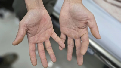
Patient NTH, 77 years old, had a history of brain tumor for 23 years, and often faced epileptic seizures. During an unfortunate epileptic seizure, the patient fell, resulting in serious facial injuries.
At E Hospital, after examination and CT scan, doctors diagnosed an extremely complicated mandibular fracture: 2 fractures, including the right horizontal branch and the opposite condyle (left side).
Many challenges were faced by the team when deciding to operate on the patient. First, the patient was elderly (77 years old) with a history of high blood pressure, diabetes, and poor health, posing many potential risks in a lengthy surgery. Meanwhile, the underlying brain tumor required simultaneous coordination.
The patient's jawbone condition was particularly difficult as he had lost all his lower teeth for many years, leading to severe jawbone atrophy, with the height of the lower jawbone being less than 2cm. This made it almost impossible to ensure accuracy and stability with traditional bone correction and fixation methods.
Faced with a difficult case, doctors from the Department of Plastic and Maxillofacial Surgery, E Hospital decided to apply personalized 3D technology to find the optimal solution.
The doctors implemented a systematic procedure including: Virtual diagnosis and planning by entering the patient's CT-scan data into specialized software. In the 3D virtual environment, the doctors performed "trial surgery", moving each jaw fracture fragment to the correct anatomical position before the injury, and repositioning the condyle to the exact position in the joint socket.

After obtaining the perfect virtual correction plan, a stereolithographic model of the patient's mandibular bone block (with the corrected bone position) was printed using 3D technology.
Based on this 3D printed model, a titanium splint for bone fixation was pre-formed to ensure an absolute fit with the bone surface after correction. The splint was then autoclaved, ready for surgery.
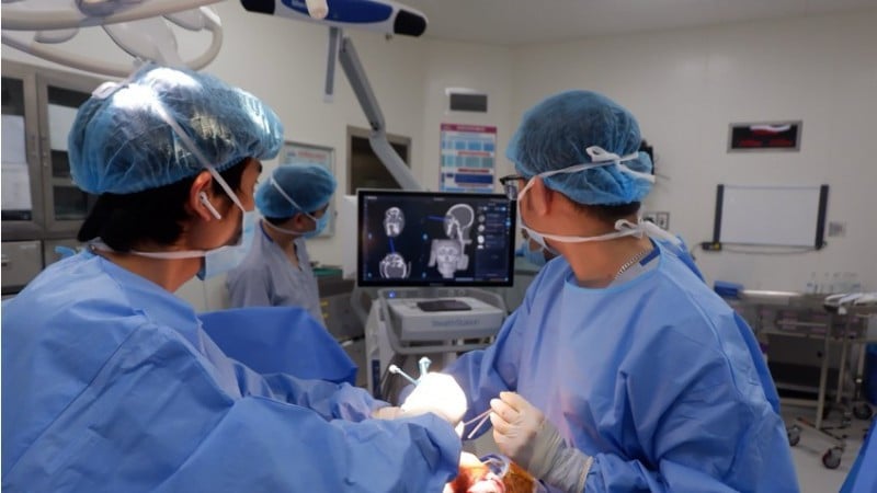
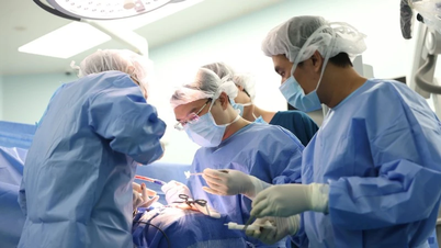
The surgery was performed with the smooth coordination of two surgical teams: Neurosurgery and Maxillofacial Surgery.
First, the Neurosurgery team applied a modern navigation system. This technology acts as a medical "Google Maps", allowing surgeons to accurately determine the tumor boundaries and healthy brain structures, thereby removing brain tumor tissue meticulously and with maximum safety.
Immediately after, the Maxillofacial Surgery team stepped in. Thanks to the "personalized" titanium splint prepared in advance, the surgeon did not waste time breaking the splint during the surgery. This splint acted as a perfect surgical guide, helping to quickly bring the broken bone fragments back to the planned position and fix them firmly.
The application of 3D technology in maxillofacial trauma surgery at E Hospital has brought outstanding benefits. Especially with this complex case, the surgery is of maximum precision, ensuring that the broken bone is returned to the correct anatomical position, restoring function and perfect aesthetics.
Pre-preparation of the splint significantly reduces the surgical time and minimizes the risk of complications for elderly patients. This method also overcomes the disadvantages of severely atrophic jawbones, where traditional techniques are difficult to apply.
Source: https://nhandan.vn/ung-dung-cong-nghe-3d-trong-phau-thuat-xuong-ham-phuc-tap-cho-benh-nhan-mac-u-nao-post918384.html

![[Photo] The 5th Patriotic Emulation Congress of the Central Inspection Commission](https://vphoto.vietnam.vn/thumb/1200x675/vietnam/resource/IMAGE/2025/10/27/1761566862838_ndo_br_1-1858-jpg.webp)


![[Photo] Prime Minister attends the 28th ASEAN-China Summit](https://vphoto.vietnam.vn/thumb/402x226/vietnam/resource/IMAGE/2025/10/28/1761624895025_image-2.jpeg)
![[Photo] National Assembly Chairman Tran Thanh Man receives Chairman of the House of Representatives of Uzbekistan Nuriddin Ismoilov](https://vphoto.vietnam.vn/thumb/1200x675/vietnam/resource/IMAGE/2025/10/27/1761542647910_bnd-2610-jpg.webp)
![[Photo] Party Committees of Central Party agencies summarize the implementation of Resolution No. 18-NQ/TW and the direction of the Party Congress](https://vphoto.vietnam.vn/thumb/1200x675/vietnam/resource/IMAGE/2025/10/27/1761545645968_ndo_br_1-jpg.webp)


Comment (0)