
Why should we have ultrasound screening for fetal abnormalities?
Birth defects occur when a fetus is developing in the womb and can affect its appearance, organ function, physical and mental development. Birth defects are one or more abnormalities in the form of physical or genetic defects that cause adverse effects on the pregnancy and the baby when born.
Major birth defects are the leading cause of death in newborns. Approximately 90% of fetal malformations cannot be identified from clinical presentation. Not all birth defects are preventable, which is whyeducation and awareness about birth defect prevention is so important.

Master - Doctor CKII Truong Quang Hung, Head of Obstetrics and Gynecology Clinic 315 Au Co branch (HCMC)
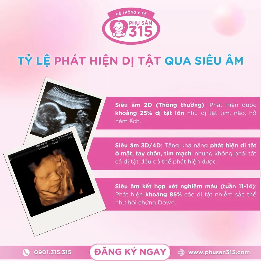
According to Master - Doctor CKII Truong Quang Hung, Head of Obstetrics and Gynecology Clinic 315 Au Co branch (HCMC): ultrasound combined with other tests is an easy-to-perform method, relatively safe, reasonable cost and has accurate results from 85-90% of fetal abnormalities to promptly detect Down syndrome, congenital defects, risk of chromosomal abnormalities... However, accuracy depends a lot on the equipment system and expertise of the examining doctor.
Combining ultrasound and maternal blood analysis in first trimester screening

Obstetrics 315 has modern ultrasound technology, clear images - so mothers can clearly see every movement of their baby. Here, pregnant mothers receive individual care, doctors understand each mother's individual needs.
Nuchal translucency (NTP) is the accumulation of fluid under the skin and at the back of the fetus's neck. Based on the NTP index, doctors can predict the risk of Down syndrome and other birth defects in the fetus.
Fetal nuchal translucency varies with gestational age and the risk of fetal chromosomal abnormalities is calculated based not only on the thickness of the nuchal translucency (an ultrasound marker) but also on the mother's age and weight, and history of previous pregnancies.
To reduce the positive screening rate, it is necessary to combine it with the analysis of freeBeta hCG and PAPP-A (Double test) in maternal blood. This combination reduces the positive screening rate from 8.3% if only using ultrasound screening to 2.4% and increases the detection rate from 80% to 90%.
Is increased nuchal translucency thickness a sure sign of fetal birth defects?
315 Obstetrics and Gynecology System provides modern and convenient health care services, helping pregnant mothers enjoy every moment of pregnancy with peace of mind.
Answering this question, Master - Doctor CKII Truong Quang Hung, Head of Obstetrics Clinic 315 Au Co branch (HCMC) said that the increase in the thickness of the nuchal translucency does not mean that the fetus is abnormal, this is only an indicator or suggestion for the doctor to conduct further tests.
If the fetal nuchal translucency is >= 3mm thick with a crown-rump length (CRL) between 45 - 84 mm, the fetus will be at risk of carrying chromosomal abnormalities, congenital heart defects or other serious defects.
All pregnant women with fetal nuchal translucency >= 3mm need to be counseled on risks and perform specialized tests to evaluate fetal chromosomes and monitor fetal abnormalities via ultrasound, especially congenital heart abnormalities. About 90% of pregnant women with fetal nuchal translucency less than 4.5mm will give birth to healthy babies. About 80% of pregnant women with fetal nuchal translucency from 4.5 - 6.4mm and 45% of pregnant women with fetal nuchal translucency >= 6.5mm will give birth to healthy babies.
When do pregnant women need amniocentesis and chorionic villus sampling?

Amniotic fluid plays an important role in the development of the fetus. Amniotic fluid helps nourish the embryo, protect and shield the fetus from collisions and creates a sterile environment for the fetus to develop.
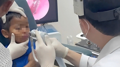
Amniocentesis is a procedure used to obtain a small sample of the amniotic fluid that surrounds the fetus. Amniocentesis is performed to diagnose chromosomal disorders and open neural tube defects (ONTDs), such as spina bifida. Furthermore, amniocentesis as a prenatal test can also detect hundreds of different genetic disorders, such as cystic fibrosis, the neurodegenerative Tay-Sachs disease, sickle cell disease, as well as neural tube defects.
It is important that the results of the amniocentesis test have a great influence on the management of pregnancy. With modern technology today, this measure is performed very easily. Amniocentesis can be performed from week 15-20 of pregnancy with a risk of increased chromosomal abnormalities.
Chorionic villus sampling (CVS) is the removal of a small amount of placental tissue from the uterus. During routine prenatal checkups or tests, if the fetus is diagnosed as having a high risk of birth defects, or if the family has a history of certain genetic diseases such as hemophilia, a chorionic villus sampling will be performed to detect genetic disorders in the fetus. The procedure is performed in the later stages of the first trimester of pregnancy, especially in the 10th to 13th week of pregnancy.
Health System 315:
Hotline: 0901.315.315
Ivy Health International Clinic https://www.ivyhealthvn.com/
315 Obstetrics and Gynecology System https://www.phusan315.com/
Children's Health System 315 https://www.nhidong315.com/
Pediatric Immunization System 315 https://www.tiemchungnhi315.com/
315 Eye Health System https://www.mat315.com/
Cardiovascular - Diabetes Healthcare System 315: https://www.timmachtieuduong315.com/
Source: https://www.sggp.org.vn/vi-sao-me-bau-khong-duoc-quen-sieu-am-tam-soat-di-tat-thai-nhi-post797915.html


Comment (0)