
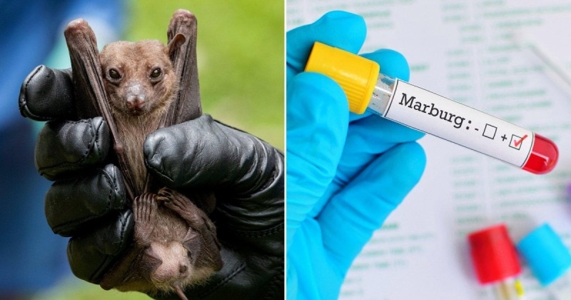
Medical news October 14: Strengthening quarantine, preventing Marburg cases from border gates
The Ministry of Health requested relevant agencies to have plans to respond to Marburg epidemic situations at each border gate with the participation and coordination of competent authorities at the border gate and local health agencies.
Strengthening quarantine to prevent Marburg cases from border gates
The Department of Preventive Medicine (Ministry of Health) said that according to information from the Infectious Disease Surveillance System, since the end of September 2024, Rwanda (Africa) has recorded the first case of Marburg disease in this country.
 |
| The Ministry of Health requested relevant agencies to have plans to respond to Marburg epidemic situations at each border gate with the participation and coordination of competent authorities at the border gate and local health agencies. |
As of October 10, authorities had recorded a total of 58 cases, including 13 deaths, in 7 of the country's 30 districts, about 70% of the cases were medical staff.
Marburg disease is a dangerous infectious disease caused by the Marburg virus. This is a virus transmitted from animals to humans, causing severe bleeding in many parts of the body. The disease is highly contagious and has a high mortality rate (50% can be up to 88%).
Currently, there is no vaccine or specific treatment for the disease. The disease is classified as group A in our country's Law on Prevention and Control of Infectious Diseases.
According to the Department of Preventive Medicine, some countries such as the United States, China, and South Korea have increased medical measures at border gates to control the entry of Marburg disease.
To proactively monitor, detect and control the Marburg epidemic entering our country, the Department of Preventive Medicine has sent an urgent document to the Institute of Hygiene and Epidemiology/Pasteur; International Health Quarantine Centers, Centers for Disease Control of provinces and cities with medical quarantine activities to update information on countries/territories that are recording Marburg cases to strengthen and proactively closely monitor subjects subject to medical quarantine from these areas entering, transiting and importing through border gates in our country.
Fully implement personal protective measures for officers, employees and people in contact with suspected/infected cases, to prevent infection among medical staff and spread to the community.
Units must prepare rooms and temporary quarantine areas for suspected and infected cases at border gates (if necessary); equipment, chemicals, and medicines must be ready for immediate use in case of an epidemic.
At the same time, training was provided to improve the capacity of medical quarantine officers in monitoring and controlling Marburg disease; attention was paid to infection prevention and control.
Continue to organize communication at border gates for passengers and people about prevention measures, especially the need to immediately notify medical facilities when they detect symptoms and epidemiological factors related to Marburg disease within 21 days from the date of entry into Vietnam.
Review and update contingency plans to respond to Marburg epidemic situations at each border gate with the participation and coordination of competent authorities at the border gate and local health agencies, including attention to accompanying medical staff, means of transport for suspected and infected people, and medical facilities that can receive care and treatment.
The Institutes of Hygiene and Epidemiology/Pasteur provide guidance, training, and support to localities on surveillance and prevention measures, sampling, and safe transportation of specimens; and receive specimens for definitive diagnosis of Marburg disease from localities.
Continue to strengthen testing capacity, diagnose Marburg disease, as well as review and strengthen rapid response teams at units, ready to respond when suspected or infected cases are recorded in localities.
According to medical experts, the Marburg virus can be transmitted from animals to humans through direct contact with bodily fluids of infected animals.
In addition, this virus is also transmitted from person to person through direct contact with the blood and secretions of an infected person or contaminated surfaces.
The incubation period ranges from 2 to 21 days, beginning with high fever, chills, severe headache, muscle pain. Around the fifth day after the onset of the disease, a maculopapular rash may appear, most prominent on the trunk (chest, back, abdomen), nausea, vomiting, chest pain, sore throat, abdominal pain and diarrhea may appear.
Symptoms become increasingly severe and may include jaundice, pancreatitis, severe weight loss, delirium, shock, liver failure, massive bleeding, and multiple organ dysfunction.
Clinical diagnosis is difficult because the disease has symptoms similar to other infectious diseases (malaria, typhoid, Ebola hemorrhagic fever, etc.). The disease has a high mortality rate (the number recorded in previous outbreaks is 24% to 88%).
According to experts, to prevent epidemics, hospitals need to have measures to detect early cases entering Vietnam through exploiting epidemiological history and clinical symptoms.
Ho Chi Minh City: The risk of Marburg disease entering is not high, but it can still happen.
The World Health Organization assesses the risk of Marburg spreading as low at the global level and recommends against imposing any travel or trade restrictions on Rwanda in light of the ongoing outbreak there.
According to the representative of the Ho Chi Minh City Department of Health, the risk of Marburg disease entering Ho Chi Minh City is not high, but it can still happen. By air, the risk of entering the City is quite low when there are no direct flights and incoming passengers are screened before leaving the country.
The possibility of maritime penetration is very low, Rwanda has only 1 maritime port in Kigali, according to vessel entry data from January 2023 to September 30, 2024, there are no vessels directly from this maritime port.
In addition, the shipping time from Africa to Ho Chi Minh City by sea usually lasts from 25-40 days, longer than the longest incubation period of Marburg (21 days).
Although the WHO has assessed the risk of this outbreak as low at the global level, some countries such as South Korea, China, and the United States have also strengthened medical measures at border gates to control the disease from entering.
On October 11, 2024, the Department of Preventive Medicine, Ministry of Health of Vietnam also issued a document directing the implementation of disease control measures at border gates. The Department of Health directed the City Center for Disease Control to strictly implement, especially monitoring passengers from flights related to Rwanda.
Faced with the ever-changing epidemic situation in the world, the Ho Chi Minh City Health Department has proactively implemented measures such as: increasing information updates on MVD as well as other emerging infectious diseases in the world;
Strengthening surveillance of people entering from epidemic areas according to the guidance of the Ministry of Health, being ready to intervene if an imported case is detected; raising awareness about risk factors for Marburg virus infection and protective measures that individuals can take are effective ways to reduce human transmission.
People should limit unnecessary travel to countries with outbreaks. For those who have traveled to countries with outbreaks, if they discover that they have symptoms of suspected illness, they should immediately go to a medical facility and provide medical staff with full information about their travel history to epidemic areas for timely diagnosis and treatment as well as to limit infection.
The Ho Chi Minh City Department of Health will continue to monitor and provide information as soon as official information is received from WHO and the Vietnamese Ministry of Health.
The City Health Department calls on people to refer to information about epidemics posted on official sources, with citations (if reposted) to avoid unverified information that causes panic and anxiety.
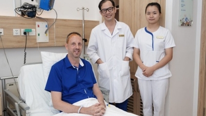
Surgery to remove giant hemangioma to save little girl's legs
The vascular tumor existed in the girl's abdominal cavity for many years, compressing the spinal cord, causing loss of feeling in both legs, and risking permanent paralysis if the tumor was not surgically removed.
Four years ago, Linh (15 years old, living in Nghe An) was diagnosed with a vascular malformation in the area next to the lumbar spine that formed a hemangioma. After four sclerotherapy treatments at a major hospital, the tumor volume has decreased, and there is no longer a risk of rupture causing bleeding. For the past two months, Linh has often had tension in her lower abdomen, and her legs are numb and she cannot move. She was taken to Tam Anh Hospital for examination.
The patient was admitted to the hospital with a very weak left leg, severe pain, and almost unable to walk. Paraclinical examination recorded two tumors in the spinal canal measuring 10x5x3 cm and 4.5x1x1 cm, and one tumor in the iliopsoas muscle measuring 10x12 cm located in the retroperitoneal space, below the kidney, behind the colon, on the left side of the spine.
Part of the tumor spread into the spinal canal, compressing the spinal cord and causing the patient's left leg to gradually weaken. In addition, the tumor also pushed the iliopsoas muscle forward, pushed the left kidney back, and displaced the ureter and colon. If the tumor was not quickly removed, the risk of the patient becoming permanently paralyzed was very high.
Specialists in diagnostic imaging, neurosurgery, thoracic-vascular surgery, vascular intervention, and urology consulted to find the most effective solution to treat hemangioma.
Determining that it was impossible to remove the entire tumor in one surgery, the team decided to perform two major surgeries: First, to release the hemangioma compressing the nerves in the spinal canal to improve the patient's walking function, then to remove the remaining large tumor in the retroperitoneal space.
To pave the way for the two major surgeries to be successful, the doctor performed a tumor embolization procedure. CT images help to accurately identify the blood vessels that feed the tumor, allowing the doctor to perform embolization to block these branches, preventing blood from reaching the tumor and helping to shrink the tumor size, while reducing the risk of blood loss during surgery.
One day later, the doctor and surgical team, with the help of K.Zeiss Kinevo 900 microscope and large 3D images, opened an incision in the back and completely removed the two tumors that had spread into the spinal canal.
The image of the lesion resembles a bunch of grapes with a fruit-like structure, each fruit is an image of a capillary bulging with blood inside. After surgery, the patient's symptoms of numbness and weakness in the legs improved significantly. Linh can walk with assistance. The pathological results confirmed that this was a cavernous hemangioma.
A week later, Dr. Nguyen Anh Dung, Head of the Department of Cardiovascular and Thoracic Surgery, Cardiovascular Center, Tam Anh General Hospital, Ho Chi Minh City, and his team performed the second surgery, opening an incision in the left side of the back, separating the remaining tumor from the surrounding tissue.
During the surgery, doctors face the risk of massive bleeding (due to the tumor being formed from excessive blood vessel proliferation) as well as damage to nearby organs. The worst case scenario is having to remove the left kidney if the hemangioma attached to this organ cannot be separated.
To prevent risks, the doctor carefully examined the CT images before surgery to determine the exact location and level of compression of the tumor. In addition, although the tumor was large, it was not too tightly attached and still had boundaries with other organs. Thanks to that, the team was able to remove the entire hemangioma after three hours, freeing the kidney, colon, ureter, and aorta from long-term compression.
A day after surgery, Linh no longer had any abdominal pain, had a good appetite, and was instructed in physical therapy to fully restore her ability to walk. The patient was discharged a week later in a healthy state, with movement restored to 4/5 of both legs.
Cavernous hemangiomas are a type of vascular malformation (other types include arteriovenous malformations, dural arteriovenous fistulas, progressive venous anomalies, and telangiectasias). Cavernous hemangiomas are clusters of abnormal blood vessels filled with blood.
The tumor may grow but is not cancerous and does not spread to other parts of the body. Most cavernous hemangiomas appear in both cerebral hemispheres, sometimes in the posterior fossa or brainstem, rarely forming in the spinal cord or peritoneal cavity like patient Linh.
According to doctors, anyone is at risk of developing cavernous hemangioma. However, the disease has a genetic factor, so if one parent has this disease, the child born has a 50% risk of developing the disease.
Patients with cavernous hemangioma need to comply with treatment as prescribed by the doctor, maintain a healthy lifestyle to improve overall health, limit complications. Post-surgery patients may need to combine physical therapy, speech therapy... to recover quickly.


![[Video] The craft of making Dong Ho folk paintings has been inscribed by UNESCO on the List of Crafts in Need of Urgent Safeguarding.](https://vphoto.vietnam.vn/thumb/402x226/vietnam/resource/IMAGE/2025/12/10/1765350246533_tranh-dong-ho-734-jpg.webp)


Comment (0)