
On the morning of September 18, doctors at Vinmec Times City International General Hospital and engineers from the Center for 3D Technology in Medicine, VinUni University announced that they had successfully performed radical surgery to remove a 11.5cm mediastinal tumor and reconstructed the patient's chest using titanium material.
Journey of chest wall reconstruction for patients with mediastinal tumors
A 55-year-old female patient ( Ha Nam ) had severe pain in her left chest for many weeks, the pain was constant, especially increased when breathing, causing difficulty in daily activities. The patient went to the provincial hospital for examination and discovered a chest tumor (anterior mediastinal tumor). The patient was transferred to Vinmec for multidisciplinary consultation and treatment plan.
The results showed that the large mediastinal tumor, up to 11.5cm, had complicatedly invaded the left chest wall, ribs 2, 3, 4, the upper lobe of the left lung and part of the sternum, causing serious compression on the heart, lungs and surrounding organs.
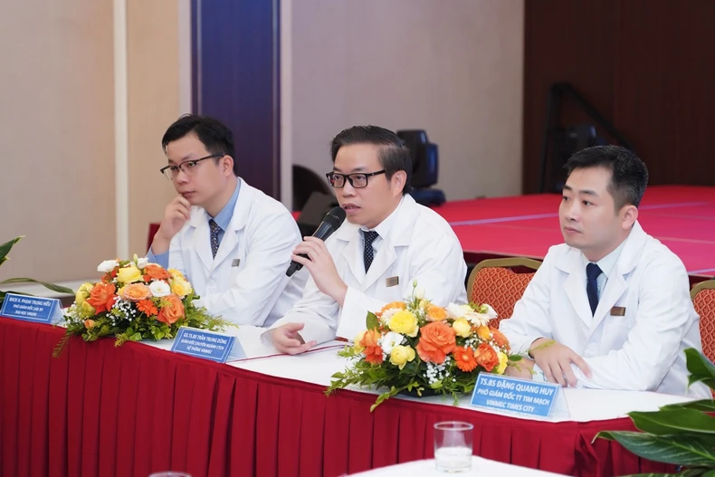
Dr. Dang Quang Huy (Deputy Director of the Cardiovascular Center, Vinmec Times City) said that the case was diagnosed at a late stage, chemotherapy and radiotherapy were no longer effective, and could only be resolved by wide surgical excision of the tumor along with the sternum and adjacent ribs.
In addition to eradicating the tumor, the case also posed a major challenge in reconstructing the chest wall to protect the heart and lung function after surgery. If not properly reconstructed, the risk of respiratory failure and internal organ injury will increase significantly.
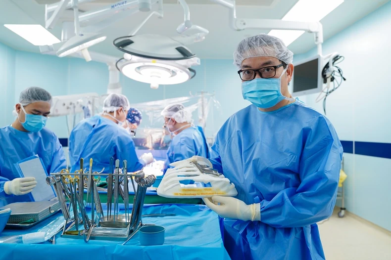 |
Professor, Dr. Tran Trung Dung (Director of Orthopedic and Musculoskeletal Trauma, Vinmec Healthcare System) and 3D printing technology products before transplant surgery for patients. |
Previously in Vietnam in particular and Southeast Asia in general, large thoracic defects after cancer surgery were often covered by using muscle-skin flaps from other locations, creating a large scar. The artificial materials used in the past were only for covering the shape, not protecting the heart and lungs inside the thoracic cavity in the correct position and functioning normally, and against external impacts.
“With the old technique, patients had to transfer the skin and muscle to the abdominal and back skin, causing blood loss, psychological trauma, severe damage, and a recovery time of 2-3 weeks. Reconstructing the chest wall is a challenge to ensure effective coverage, avoid trauma, and ensure recovery and maintain respiratory function for the patient. Therefore, traditional surgical options cannot be considered the optimal solution,” said Dr. Huy.
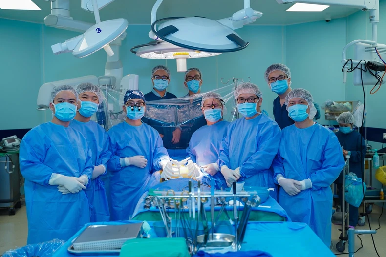 |
Surgical team before performing transplant on patient. |
The Cardiovascular Center's assignment for the 3D LAB room at VinUni University was to ensure three factors: anatomically, the chest must fit snugly; ensure respiratory function for the patient; and ensure there is a mesh to prevent lung hernia.
Specialist Doctor II Pham Trung Hieu, Deputy Director of LAB 3D, VinUni University, said that the engineering team was very thoughtful when receiving the request. “We have studied many world medical literatures and found that there is no method that can ensure all three factors. We have to solve each problem in turn,” said Mr. Hieu.
First, the LAB staff created a printed layer that was elastic but fixed to the bone, creating a mesh to prevent lung herniation. Next, instead of having to wait 6-12 weeks for the material to be ordered overseas and not being sure if it would fit the patient's body, the staff designed a chest reconstruction material that fit the patient.
Finally, the implantation process is precisely calculated by performing the first simulation on the computer, the second on the model, and the third when the final design is available before surgery is performed.
 |
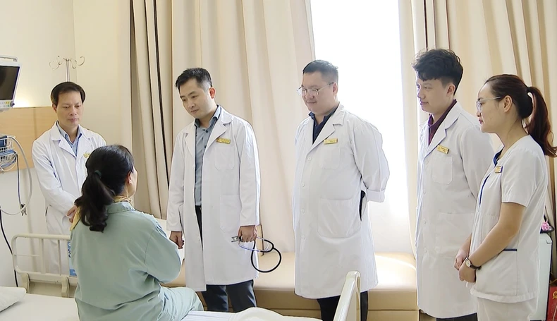
Doctors share about the surgical process with patients. |
To solve this problem, the design team, including Vinmec cardiothoracic and orthopedic trauma experts and the engineering team from the Center for 3D Technology in Medicine, VinUni University, spent nearly three weeks tirelessly researching and improving to overcome the limitations of previous designs, testing dozens of simulation scenarios to achieve meticulousness and sophistication in design, ensuring thinness and precision according to the highest standards.
The product is not only a breakthrough in design but also integrates an anti-lung hernia mesh, surpassing surgeries around the world that require printing many separate pieces to protect the lungs and heart. This innovation helps to minimize the risk of separate pieces being displaced in the body after surgery.
The entire process from design to production is approved by the Professional Council and Ethics Council of Vinmec Healthcare System, in close coordination with VinUni Institute of Health Sciences, ensuring the surgery is successful with optimal quality.
“This is a superior design, the first time it has been done in Southeast Asia. This material has great biocompatibility and can last a lifetime in the patient’s body. With this new technique, the patient goes back to the old incision, the dissection is gentle, only exposing the ribs and sternum, then the doctors proceed to insert the complete graft into the patient’s body with screws. After 5 days of surgery, the patient can walk and live normally,” said Dr. Hieu.
Three hours of total left breast reconstruction for patient
On September 11, under the direction of Dr. Dang Quang Huy (Deputy Director of Cardiovascular Center, Vinmec Times City) and Professor, Dr. Tran Trung Dung (Director of Orthopedic and Musculoskeletal Trauma, Vinmec Healthcare System), the surgery to reconstruct almost the entire left chest for the patient was successful after nearly 3 hours.
Previously, the patient had undergone the first surgery to completely remove the mediastinal tumor that invaded the chest area. In this second surgery, the doctors focused on restoring the anatomical structure of the chest area, ensuring respiratory function and the necessary protection for important internal organs.
 |
The patient recovered after surgery. |
During the surgery, the implant was tested for functionality. The fit was 99% and the patient was able to breathe normally. Just one day after surgery, the patient was able to sit up and talk normally, recovered well and was discharged from the hospital after only five days.
The precision of the surgery thanks to 3D technology, along with advanced pain relief techniques ESP, has shortened the surgery time and helped patients recover quickly, with the hospital stay reduced by only half compared to normal.
According to published scientific reports, in the past 10 years, there have been about 50 cases of 3D thoracic cavity reconstruction using titanium alloy materials in the US, Europe, Korea, and China. Notably, the patient at Vinmec is the first case in Southeast Asia and Vietnam is the fourth country in Asia to use all artificial bones and anti-hernia mesh for the heart and lungs that are completely designed and 3D printed from titanium material, without integrating or mixing other materials.
Professor, Dr. Tran Trung Dung (Director of Orthopedic and Musculoskeletal Traumatology, Vinmec Healthcare System) was very excited after this surgery when these results were highly appreciated by friends at home and abroad. This is a breakthrough technique, helping patients undergo only a light surgery, ensuring a quick recovery for the patient, maintaining very good respiratory function.
According to Professor Dung, nearly 3 years ago, Vietnam also performed a chest wall reconstruction using the old method, but the patient did not survive more than 2 years because of the effects on heart and lung function.
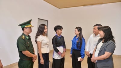
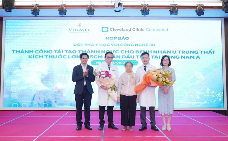 |
| The management congratulated the patient on his full recovery and discharge from the hospital. |
The success of the surgery not only created a turning point in the field of large chest defect restoration in Vietnam but also opened up great opportunities in other fields, such as maxillofacial defect reconstruction, soft tissue reconstruction, and precise stent intervention, contributing to improving treatment effectiveness and shortening recovery time for patients.
According to Dr. Dang Quang Huy, Deputy Director of the Cardiothoracic Center, the application of 3D printing technology in the cardiovascular field in particular is a trend in the world and some countries in the US and Europe are piloting initial application research.
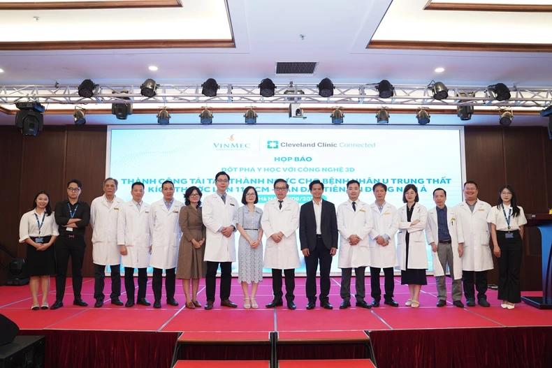 |
The team of doctors and medical staff congratulated the success of the surgery. |
With this achievement, doctors at the Cardiovascular Center have more confidence and experience to have the best intervention and treatment plan for patients with similar diseases. “This is our long-term direction. We will continue to work more with VinUni Center, keeping up with world trends,” Dr. Huy shared.


Comment (0)