
The effectiveness of LAMS technique
Can Tho Central General Hospital recently successfully treated a 29-year-old male patient, PTN, residing in Can Tho City, who suffered from a large pseudopancreatic cyst due to chronic pancreatitis. The patient was admitted at 3:20 PM on November 17th with mild abdominal distension, dull abdominal pain, accompanied by vomiting and frequent diarrhea. Due to the recurring illness over many years, the patient's health had deteriorated, his physical condition was weak, his appetite was poor, and he had lost a significant amount of weight.

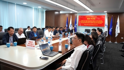
The interventional medical team is performing an endoscopic ultrasound on the patient.
Contrast-enhanced abdominal CT scan revealed a multi-lobed cystic fluid collection in the posterior gastric region, accompanied by diffuse thickening of the colon wall and dilated loops of the small intestine. The patient was admitted to the General Surgery department for comprehensive evaluation and intensive medical treatment before intervention. Following general treatment, the patient underwent upper gastrointestinal endoscopy on November 25th. The examination revealed a pseudopancreatic cyst located in the pancreatic tail, adjacent to the gastric wall, providing favorable conditions for endoscopic drainage.
Through interdisciplinary consultations, the medical team unanimously chose the LAMS stent placement method to create a connection between the cyst and the stomach, allowing for safe drainage of pancreatic fluid. This is a minimally invasive technique that is highly effective for complex pancreatic cystic lesions.
On December 3rd, the intervention was performed under endoscopic ultrasound guidance. During the examination with a linear probe, the doctor discovered a cyst measuring 9x11cm with thick walls and containing a significant amount of fluid. First, a 19G aspiration needle was inserted through the stomach wall into the cyst to create an access pathway. After the guidewire was correctly positioned, the doctor used a cutting instrument to widen the cyst wall. Next, a LAMS stent was threaded along the guidewire, positioned, and expanded to create a permanent connection between the cyst and the stomach. Once the stent opened, a steady flow of lemon-yellow fluid was observed; a portion of the fluid was collected for testing to assess for infection or related complications.
The procedure lasted approximately 20 minutes. Thanks to the self-expanding properties and flared ends of the LAMS stent, the drainage pathway remained stable, ensuring continuous fluid drainage, minimizing the risk of leakage, and reducing potential complications. By the morning of December 9th, the patient was alert, vital signs were stable, the abdomen was soft, pain had significantly decreased, and there were no longer any signs of resistance. The patient continues to be monitored in the General Surgery department and is expected to be discharged in the next few days.
Expanding access to modern treatment options for patients.
According to Dr. Nguyen Khac Nam, Deputy Head of the General Surgery Department at Can Tho Central General Hospital, pancreatic pseudocysts are a common complication after acute or chronic pancreatitis, forming when pancreatic fluid leaks out of the pancreatic duct and accumulates in the surrounding tissue. Over time, the affected area is surrounded by a layer of necrotic fibrous tissue, forming a cyst, but without the epithelial layer found in true pancreatic cysts. Cysts can form in just a few days, or sometimes take many months, usually located in the body or tail of the pancreas, or spreading to the posterior stomach and colonic mesentery.

The patient was stable after the intervention.
Symptoms are very diverse: persistent epigastric pain, nausea, weight loss, gastrointestinal compression, digestive disorders, or fever and jaundice when the cyst puts pressure on the bile ducts. If not treated promptly, the cyst can cause infection, necrosis, rupture into the abdominal cavity, or internal bleeding, all of which are life-threatening complications.
Previously, open surgical or percutaneous drainage methods were often applied, but the degree of invasion was high, recovery time was long and there were many potential risks. Internal drainage by surgery connecting the cyst to the stomach or small intestine also required high technique and prolonged hospital stay.
Dr. Nguyen Thi Quynh Mai, Head of the Endoscopy Department at the hospital, stated that endoscopic ultrasound (EUS) combined with LAMS stent placement is a significant breakthrough in the treatment of pancreatic pseudocysts. EUS allows doctors to access the cyst with high precision, clearly observe tissue structure, and avoid damage to blood vessels or adjacent organs. LAMS stents create a wide and stable passage, facilitating rapid fluid drainage, reducing treatment time, and minimizing complications.
The success of the first case in the Mekong Delta demonstrates the growing capacity of Can Tho Central General Hospital to implement modern interventional techniques. Proactively mastering LAMS technology allows patients in the region to access advanced treatment methods previously only available at major centers in Ho Chi Minh City or Hanoi .
Dr. Mai emphasized that, in the context of the increasing demand for treatment of pancreatic and biliary diseases, the implementation of EUS-LAMS technology not only helps reduce the burden of surgery but also improves the quality of treatment, shortens recovery time, and significantly improves the quality of life for patients. This success also paves the way for the hospital to continue applying more minimally invasive techniques in the future.
Source: https://doanhnghiepvn.vn/tin-uc/dong-bang-song-cuu-long-lan-dau-dung-stent-lams-dieu-tri-nang-gia-tuy/20251209032603551


![[Video] The craft of making Dong Ho folk paintings has been inscribed by UNESCO on the List of Crafts in Need of Urgent Safeguarding.](https://vphoto.vietnam.vn/thumb/402x226/vietnam/resource/IMAGE/2025/12/10/1765350246533_tranh-dong-ho-734-jpg.webp)


Comment (0)