
(CPV) – Recently, the Department of Maxillofacial Surgery (B8) – Military Hospital 175 has successfully performed for the first time a surgery to resect the lower jaw bone and reconstruct it with a free fibula musculocutaneous flap, applying modern 3D technology. This is a breakthrough in the treatment of ameloblastoma, bringing results beyond expectations in both function and aesthetics for the patient.
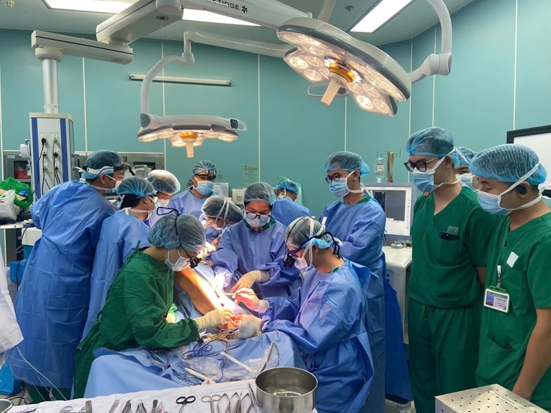 |
| 3D printing technology has been applied throughout the entire process from simulation design to surgical planning. |
Ameloblastoma is a benign tumor but has the ability to destroy bone and has the potential to become malignant if not treated promptly. Surgery to remove the jawbone to remove the tumor often causes serious functional and aesthetic losses, greatly affecting the patient's quality of life, especially in young people.
To optimize treatment results, 3D printing technology has been applied in the entire process from simulation design to surgical planning. This technology allows for precise design of bone cutting trays and reconstruction splints, jaw models and surgical guides with high precision, ensuring the procedure is performed effectively, safely and optimizes treatment results.
Regarding the specific case that was just operated on at the Hospital, male patient NQN (1995, Quang Ngai ) discovered a tumor in the lower jaw bone 6 years ago but stopped treatment after the initial monitoring period. Recently, the swelling increased, the patient came to Military Hospital 175 with a swollen lower jaw, mild red inflammation of the mucosa, and little pain. The results of the diagnostic imaging showed that the tumor destroyed the bone from tooth number 44 to the ascending branch of the right lower jaw.
The patient underwent surgery to resect the lower jaw, remove the tumor with a 1cm safe margin, and reconstruct the lower jaw with a free peroneal muscle flap. This is a complex technique, requiring skill, meticulousness, and endurance from the surgical team. However, this method is supported by 3D technology, helping doctors accurately simulate each cutting, grafting, and reconstruction operation. Thanks to that, the surgical results are optimized in both function and aesthetics, bringing outstanding efficiency to the patient.
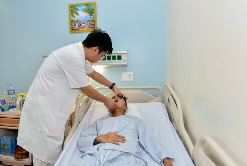 |
| After 10 days of surgery, the patient recovered well. |
The implementation process includes: The patient is designed with a cutting line and surgical guide to cut the jaw bone and fibula (cutting guide) on the software, print the guide, print a 3D jaw model after simulating bone cutting and reconstruction. Bend the splint according to the simulated jaw model. Then, cut the lower jaw bone from the high branch to the distal side of tooth 42, the fibula muscle flap after dissection will be cut according to the cutting guide. Combine the fibula into the pre-bent reconstruction splint, then combine the flap complex, splint into the lower jaw bone to ensure the bite and mobility of the jaw bone. Finally, the microsurgical technique of suturing blood vessels ensures the survival of the reconstruction flap.
After 10 days of surgery, the patient recovered well: the whole body was stable, no fever, normal vital signs, good fibula flap, dry surgical wound, fast healing, eating, chewing, opening and closing movements and facial aesthetics were significantly improved. Having a perfect 3D shaping plan will shorten the surgery time, helping the patient recover quickly.
The lower jaw bone not only plays an important role in chewing, speaking, and swallowing, but also shapes the facial structure. Therefore, reconstructing large defects after surgery is always a top priority. The free fibula flap with 3D printing technology offers many advantages: Ensuring enough bone supply for large segment reconstruction, allowing two surgical teams to perform simultaneously to shorten the surgical time while increasing the accuracy and effectiveness of treatment.
Doctor Do Van Tu - Department of Maxillofacial Surgery, Military Hospital 175, who directly performed the surgery, said: "Applying software to create "cutting guides" and print jaw models helps surgeons have the most precise cutting lines, especially helping to create the most perfect lower jaw bone from the fibula from every angle. Calculating the details of the cuts and grafts will help doctors perform surgery simply and accurately, instead of having to wait until the patient is on the operating table to perform time-consuming calculations like before. Planning and implementing the plan when applying 3D technology in microsurgery is an important factor to ensure optimal aesthetic and functional results for the patient."
“Currently, 3D technology is increasingly applied in the field of Dentistry and Maxillofacial Surgery such as orthopedic surgery, dental implant surgery, plastic surgery, etc. Therefore, using 3D technology to design simulations and plan jawbone reconstruction surgery using microsurgical fibula flaps helps to simplify, not only help improve accuracy and shorten surgery time, but also optimize treatment results, especially in complex cases such as mandibular reconstruction. The combination of 3D printing technology and microsurgical techniques is a testament to the continuous efforts of Military Hospital 175 in applying advanced technology, providing optimal solutions, meeting treatment needs and improving the quality of life for patients" - Dr. Tu further emphasized./.


![[Photo] National Assembly Chairman Tran Thanh Man attends the VinFuture 2025 Award Ceremony](/_next/image?url=https%3A%2F%2Fvphoto.vietnam.vn%2Fthumb%2F1200x675%2Fvietnam%2Fresource%2FIMAGE%2F2025%2F12%2F05%2F1764951162416_2628509768338816493-6995-jpg.webp&w=3840&q=75)

![[Photo] 60th Anniversary of the Founding of the Vietnam Association of Photographic Artists](/_next/image?url=https%3A%2F%2Fvphoto.vietnam.vn%2Fthumb%2F1200x675%2Fvietnam%2Fresource%2FIMAGE%2F2025%2F12%2F05%2F1764935864512_a1-bnd-0841-9740-jpg.webp&w=3840&q=75)


Comment (0)