
Mr. CK (61 years old, Cambodian nationality) was severely injured when a lawnmower struck him while he was working, cutting off a large chunk of his right leg and causing significant bleeding.
From Cambodia to Ho Chi Minh City seeking a better life.
Following the accident, the patient received temporary first aid in Cambodia, and was then transferred to Nam Saigon International General Hospital (Ho Chi Minh City) that same night.
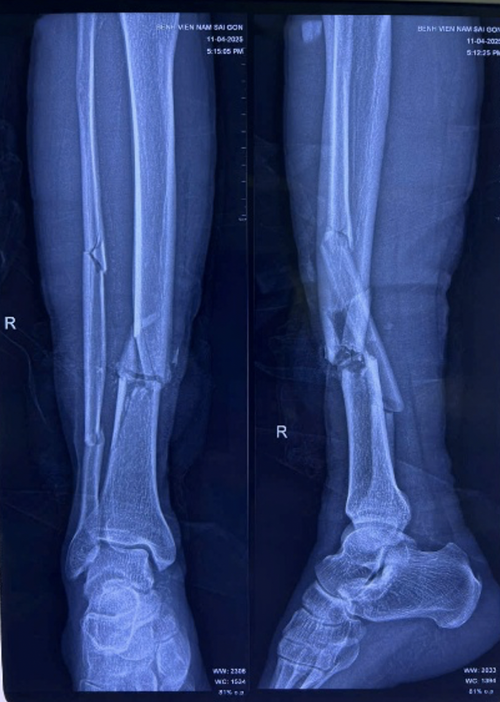
X-ray of the patient's leg before surgery (Photo: Provided by the hospital).
Dr. Son Tan Ngoc, the doctor directly treating Mr. K., said that he was admitted to the emergency room with a severe injury to his right leg, a large open wound, deformity, blood loss, and rupture of blood vessels, nerves, and tendons in the anterior tibial muscle—important structures in the foot.
The patient was diagnosed with a severe open fracture. Without timely and proper intervention, the man was at high risk of losing mobility in his foot, and might even require amputation to save his life.
The emergency team in the Orthopedics Department took advantage of the "golden hour" within the first 6 hours to perform the surgery.
Doctors clean and debride the wound, removing damaged tissue to prevent infection, and place an external fixation frame to stabilize the fractured bone – a technique that helps stabilize the bone structure without deep intervention into the injured area, facilitating the healing process.
Simultaneously, the patient underwent suturing of the anterior tibial artery, nerve, and tendon to restore blood circulation and regain sensation and motor function in the leg in the future.
The key to the surgery is the microsurgical technique of reconnecting the anterior tibial artery and nerve, requiring a highly skilled surgeon, meticulous attention to detail, and the support of specialized, modern equipment.
"We use a high-magnification surgical microscope to reconnect blood vessels with a diameter of only 1-2mm. This is a crucial factor in determining whether the severed foot will receive blood supply and be preserved," explained Dr. Son Tan Ngoc.
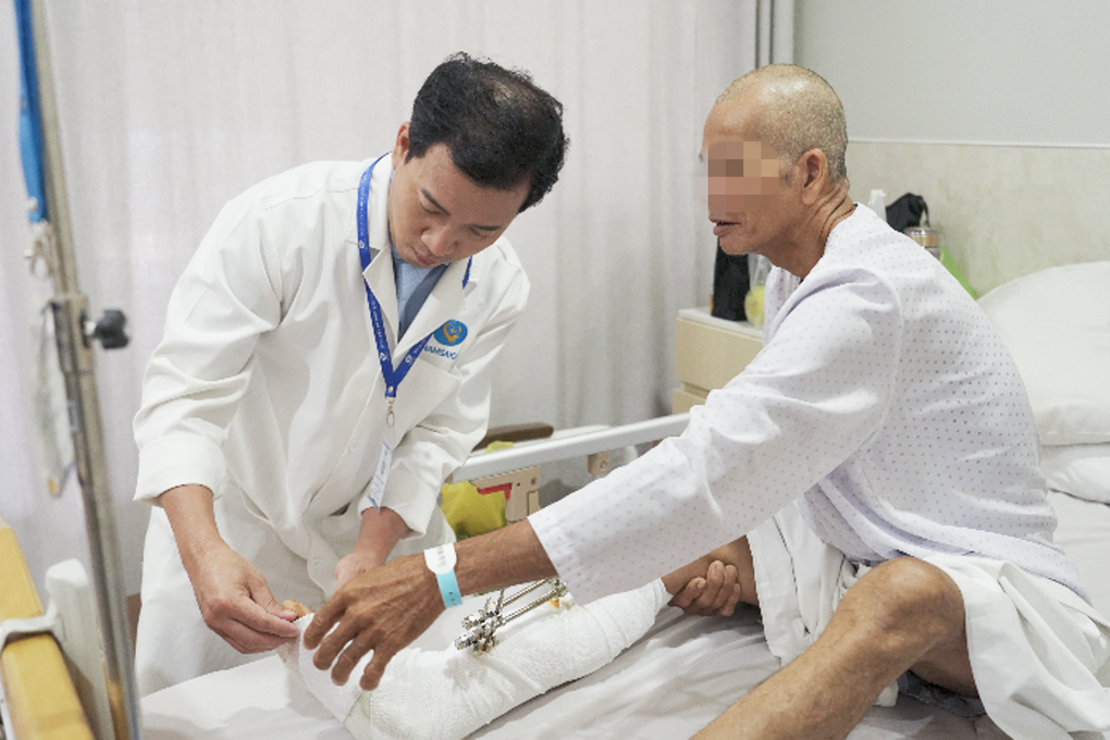
Doctors check the condition of the patient's leg after surgery (Photo: Provided by the hospital).
After more than 3 hours of surgery, the medical team successfully performed microsurgical anastomosis of blood vessels, nerves, tendons, and muscles, maximizing limb preservation for the patient, maintaining leg length, ensuring blood circulation, and restoring sensation and motor function.
Anesthesia and resuscitation also play a crucial role in ensuring the patient remains stable throughout the surgery.
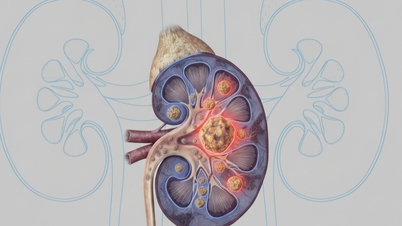
Detecting potential kidney damage.
This case didn't just involve surgery to save the foot. During a thorough examination of the patient's overall condition, doctors discovered that Mr. K. had signs of kidney stones in his right kidney due to stenosis at the ureteropelvic junction.
This condition prevents urine from draining from the kidneys to the bladder, leading to fluid retention and increasing the risk of infection, kidney tissue damage, and kidney failure if not detected and treated promptly.
Just three days after the microsurgery to reconnect the lower limb artery – when the patient's health had stabilized – the surgical team led by Dr. Le Van Hieu Nhan (specializing in Urology) performed the next surgery to reconstruct the ureteropelvic junction and remove kidney stones.
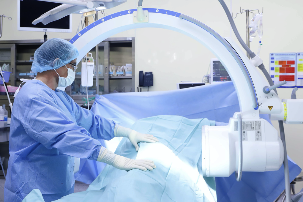
Surgery to remove kidney stones for a patient (Photo: Provided by the hospital).
The Urology team chose the intrasinus pyelotomy method to remove the stone, with a 10cm skin incision in the rib cage. This technique allows for precise access to the stone's location while thoroughly addressing the cause of the obstruction, which is the narrowing of the ureteropelvic junction.
Following the surgery, the 1.4mm kidney stone was successfully removed. The patient's ureteropelvic junction was reconstructed, ensuring smooth urine flow from the kidney to the bladder, thereby preventing the risk of stone recurrence and protecting kidney function in the long term.
"Early detection and simultaneous treatment of both issues at the appropriate time not only helps patients achieve complete recovery, but also avoids dangerous, insidious complications that may go undetected for years," Dr. Nhan shared.
After approximately two months of intensive treatment and four comprehensive surgeries, the patient's condition has now stabilized significantly, and post-operative monitoring indicators all show positive results.
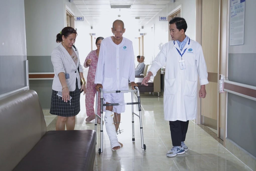
Doctors monitor and guide patients through physical therapy exercises (Photo: Provided by the hospital).
The patient's foot has shown good signs of recovery, the wound is dry and clean, with no signs of infection. Meanwhile, kidney function has improved with good urine flow and no more water retention.
According to the doctors, the above case is a prime example of the important role of the multidisciplinary collaboration model at Nam Saigon International General Hospital.
If the focus is solely on treating the initial injury while ignoring other symptoms, the patient may miss the opportunity to detect and treat dangerous underlying conditions such as ureteral obstruction, kidney stones, or impaired kidney function.
"This is also the treatment approach that the hospital always pursues: Comprehensive - Individualized - For the long-term benefit of the patient. With a team of experts and advanced equipment, the hospital not only meets the high-quality medical needs of domestic people but is also ready to receive international patients," a representative of Nam Saigon International General Hospital affirmed.
Source: https://dantri.com.vn/suc-khoe/bac-si-viet-cuu-benh-nhan-campuchia-bi-may-cat-co-chem-vao-chan-nguy-kich-20250805165235636.htm

![[Photo] Prime Minister Pham Minh Chinh holds a phone call with the CEO of Russia's Rosatom Corporation.](/_next/image?url=https%3A%2F%2Fvphoto.vietnam.vn%2Fthumb%2F1200x675%2Fvietnam%2Fresource%2FIMAGE%2F2025%2F12%2F11%2F1765464552365_dsc-5295-jpg.webp&w=3840&q=75)
![[Photo] Closing Ceremony of the 10th Session of the 15th National Assembly](/_next/image?url=https%3A%2F%2Fvphoto.vietnam.vn%2Fthumb%2F1200x675%2Fvietnam%2Fresource%2FIMAGE%2F2025%2F12%2F11%2F1765448959967_image-1437-jpg.webp&w=3840&q=75)


![[OFFICIAL] MISA GROUP ANNOUNCES ITS PIONEERING BRAND POSITIONING IN BUILDING AGENTIC AI FOR BUSINESSES, HOUSEHOLDS, AND THE GOVERNMENT](https://vphoto.vietnam.vn/thumb/402x226/vietnam/resource/IMAGE/2025/12/11/1765444754256_agentic-ai_postfb-scaled.png)


Comment (0)