
This type of tumor can contain various tissues such as hair, teeth, nails, glandular tissue, or nerve tissue and can easily cause ovarian torsion, necrosis, compression of the intestines, stomach, and blood vessels, or even rupture leading to peritonitis, severely affecting future fertility. Thanks to modern techniques and close professional collaboration, the medical team successfully removed the tumor while preserving the patient's reproductive function as much as possible in the future.
Ovarian teratoma over 20cm in a 9-year-old girl: A rare and unusual case.
According to the family, recently, 9-year-old NTKN from Hanoi has been experiencing unusual abdominal pain, accompanied by a heavy, aching sensation in the lower abdomen. In particular, her lower abdomen has noticeably swollen and shifted significantly to the right, causing the family great concern.
The family took the child to several medical facilities, and all doctors diagnosed a large localized tumor in the lower abdomen, suspected to originate from the ovary. Ultrasound and multi-slice computed tomography (MSCT) scans confirmed the presence of a mature ovarian teratoma in the abdominal cavity, exceeding 20 cm in diameter, almost completely filling the abdominal space and compressing several adjacent organs. The family researched and chose Phenikaa University Hospital for treatment, hoping the child would receive the safest and most optimal intervention.

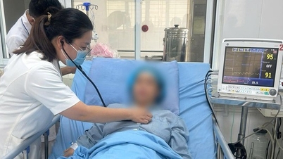
Baby NTKN was admitted to the hospital in a weak and emaciated condition, with one side of her abdomen distended, and experiencing difficulty with bowel movements and urination.
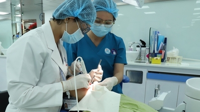
Dr. Phung Quang Thuy, Deputy Head of the Obstetrics and Gynecology Department at Phenikaa University Hospital, who directly examined and performed surgery on baby N., said this is a rare case in young children. "With tumors of such large size, the associated risks such as torsion, necrosis, and even suspected malignancy increase. The tumor is compressing many organs, leading to difficulties with bowel and bladder function and a very thin physique , " the doctor shared.
According to Dr. Thuy, ovarian teratomas in young children are a type of germ cell tumor that often does not present with typical clinical symptoms as in adults. The tumor may contain various types of tissue such as hair, teeth, nails, glandular tissue, or nerve tissue. When they grow too large, they can cause ovarian torsion leading to necrosis, compress the intestines, stomach, or blood vessels, or rupture the tumor causing peritonitis. "These risks not only threaten immediate health but also have long-term effects on the child's fertility if not treated early , " Dr. Thuy emphasized.
The surgery was successful, preserving the maximum possible reproductive capacity for the young patient.
Faced with the "enormous" size of the tumor, the medical team held a consultation and unanimously chose open surgery to ensure the child's safety. Dr. Phung Quang Thuy stated: "The biggest goal of the surgery was not just to remove the tumor, but to preserve the healthy, fragile part of the child's ovary to maintain her future fertility."

The tumor, measuring 20cm in diameter, was successfully removed while preserving as much healthy ovarian tissue as possible for the child.
The doctors carefully dissected the tissue layer by layer to avoid the risk of rupture and minimize damage to surrounding structures. After two hours of intervention, the tumor was removed intact. During the surgery, the team performed a biopsy of the tumor. The results showed a grade 1 teratoma, a benign tumor that was not a cause for concern. Dr. Thuy also added: "The surgery was successful, preserving almost all of the remaining healthy tissue of the left ovary for the child. The right ovary was also thoroughly checked and is functioning completely normally. This confirms that the child's fertility will be well maintained in the future . "

The tumor contained fat and teeth inside, and the biopsy results were benign.
N.'s mother shared emotionally: "After only 6 days of treatment, my child recovered quickly. She can walk easily, eats normally, and is in better spirits every day. My family is truly grateful to the doctors."
PhenikaaMec Obstetrics and Gynecology Department: Providing women's healthcare with a comprehensive, internationally standardized model.
The Obstetrics and Gynecology Department of Phenikaa University Hospital (PhenikaaMec) is oriented towards being a comprehensive obstetrics and gynecology center with a system of obstetric examination rooms – specialized ultrasound rooms – procedure rooms, post-procedure rooms – inpatient rooms, etc., meeting international standards.

MSc. Dr. Phung Quang Thuy - Deputy Head of the Obstetrics Department (in the middle) with experienced doctors and nurses.
The Obstetrics and Gynecology Department is also equipped with modern medical equipment such as GE Healthcare Voluson E8, E10, S21, E22 ultrasound machines, and a Signa Prime 1.5 Tesla MRI machine, providing clear images and superior diagnostic accuracy, supporting effective diagnosis and treatment of obstetric and gynecological cases. Along with a highly qualified team of professionals trained both domestically and internationally, PhenikaaMec's Obstetrics and Gynecology Department is a reputable address that helps women feel secure in caring for their reproductive health at all stages.
To learn more about PhenikaaMec Gynecology Department and its women's health services, please contact:
Phenikaa University Hospital
• Address: Group 5, Hoè Thị, Xuân Phương Ward, Hanoi City
• Website: https://phenikaamec.com/
• Hotline: 1900 886648 (free consultation 24/7)
Phenikaa University Hospital
Source: https://suckhoedoisong.vn/phenikaa-phau-thuat-thanh-cong-cho-be-gai-9-tuoi-mac-u-quai-buong-trung-20-cm-bao-toan-kha-nang-sinh-san-169251210163025554.htm


![[Video] The craft of making Dong Ho folk paintings has been inscribed by UNESCO on the List of Crafts in Need of Urgent Safeguarding.](https://vphoto.vietnam.vn/thumb/402x226/vietnam/resource/IMAGE/2025/12/10/1765350246533_tranh-dong-ho-734-jpg.webp)


Comment (0)